|
|
Know about ENT
(Otolaryngology) |
ENT Services at SCEH |
|
Ear, Nose and
Throat(ENT) Examination |
ENT diagnostic &
treatment units |
|
ENT Doctors in New York
and New Jersey |
�� |
|
ENT doctors release
national guideline on treatment for common cause of
dizziness |
|
[A prospective study of
ENT complication following surgery of the cervical
spine by the anterior approach (preliminary
results)] |
|
| �� |
|
[A prospective study of
ENT complication following surgery of the cervical
spine by the anterior approach (preliminary
results)] |
| In order to evaluate complications due to
cervical spine surgery using the anterior cervical
approach a prospective study was conducted on 125
patients. ENT Exam Unit (ENT Examination Unit) examination with the fibroscope was
employed for all the patients before the procedure.
The patients were operated on under general
anesthesia and were intubated with an armoured tube,
and then were placed in an intensive care unit for
24 hours. Assessment of deglutition and an ENT Exam Unit were performed the day after surgery.
Before surgery, two cases of vocal cord paralysis
were noted. 111 patients (88.8%) presented with
subjective disorders: problems such as sore throat, odynophagia, dysphagia, dysphagia with overspill and
hoarseness were respectively noted in 55 (44%), 34
(27.2%), 32 (25.6%), 11 (8.8%) and 13 (10.4%) cases.
Dyspnoea was found in 2 cases (1.6%). 117 patients
(93.6%) presented postoperative anomalies which were
found on the posterolateral pharyngeal wall, on the
arytenoids and on posterior third of the vocal
cords. Inflammatory and/or swollen lesions were
slight, moderate, significant or very significant in
respectively 22.4%, 22.4%, 15.2% and 1.6% of cases.
Very significant circumferential swelling of the
pharyngeal wall and of the arytenoids was
responsible for two cases of respiratory distress,
and the patients required reintubation and return to
theatre. Severe pharyngeal lesion correlated with
duration of surgery (r = 0.20; p < 0.05), with the
number levels of fusion (r = 0.02; p < 0.02) and
with the age of the patient (p < 0.02). Six patients
presented problems of mobility of the vocal cords: 3
had a right vocal cord paresis which was temporary
and 3 had paralysis, also on the right but which
persisted. There were no other complications. It is
concluded that (i) ENT complications are frequently
found in postoperative cervical spine surgery using
the anterior cervical approach, some of them being
severe. An ENT examination must be performed before
the procedure for legal reasons. It is also
recommended in the postoperative period in the case
of discomfort; (ii) patients need to be placed in an
intensive care unit during for the first 24 hours
(iii). This study needs to be attended over more
patients (iv) comparison with a control group of
patients having non cervical surgery and intubated
in the same way is needed to differentiate lesions
related to surgery or intubation. |
| Article Source: |
| http://www.ncbi.nlm.nih.gov/pubmed/9770050 |
|
Back To Top |
| �� |
|
Know about
ENT
(Otolaryngology) |
What is ENT (Otolaryngology)?
Otolaryngology is a medical specialty that deals in
the medical and surgical management and treatment of
patients with diseases and disorders of the ear,
nose, throat (ENT,ENT Examination Unit), and related structures of the
head and neck.
The special skills in ENT include diagnosis and
management of diseases of the sinuses, larynx (voice
box), oral cavity, and upper pharynx (mouth and
throat), as well as structures of the neck and face.
Otolaryngologists diagnose, treat, and manage
specialty-specific disorders of ENT as well as many
primary care problems in both children and adults.
Major problems that require medical attention
EAR
��Hole
in eardrum or CSOM (Chronic Suppurative Otitis
Media) safe and unsafe
��Hearing
Loss
��Ear
infections
��Ear
noise or ringing in the ear (Tinnitus)
��Balance
disorders / Giddiness / Vertigo
NOSE
��Allergy
/ Polyps or mass in nose
��Sinusitis
��Deviated
Septum or the nasal bone
��Snoring
��Nose
Bleeding
��Stuffy
nose
��Loss
of smell
��Headache
THROAT
��Sore
Throat
��Tonsillitis
��Hoarseness
or other voice problems
��GERD
(Gastro Esophageal Reflux Disease)
HEAD AND NECK
��Thyroid
clinic for medical & surgical management
��Neck
masses & tumoes
��Cancer
of the voice box |
| Article Source: |
| http://www.sceh.net/ent.asp |
|
Back To Top |
| �� |
|
ENT Services at SCEH |
The department of Otolaryngology (Ear, Nose and
Throat,ENT Exam Unit) is well known for the quality of services it
offers. The department has state of the art
equipment like examination unit with inbuilt head
light, suction machine, a mirror warmer and a
syringing unit.
Operation theatres are fitted with HEPA filters and
have the facility of Carl Zeiss operating
microscope. Nasal and sinus surgery equipment has
complete world class nasal endoscopes with camera
and monitor unit. The Operation theatre has a LASER
and Radiofrequency machine for doing highly
specialized surgeries.
The hospital has a fully functioning Audiology and
Speech Therapy department. Complete Hearing
evaluation is done irrespective of the age of the
child. We have facility of BERA, a special equipment
for hearing testing of young children. Adult hearing
is tested using Pure Tone Audiometer and to further
find out the cause of hearing loss impedance
audiometry is done.
Speech Therapy for stammering disorder and
articulation defects are done on a regular basis.
Headache clinic is run on every Saturday and deals
with the holistic approach to the causes and
treatment of headache in paediatric and adult
population. Extensive work up is supported by the
relevant examination and investigations.
Sleep study is done in the hospital and patients are
given advice about medical or surgical treatment of
snoring depending upon the need. Somnoplasty/UPP is
done using Laser and Radiofrequency machine.
Paediatric ENT is our recent addition involving
specialized and focused care for children below 14
years of age
The Department of ENT at SCEH offers the following
facilities
��Ear,
Nose and Throat examination in the OPD using
treatment unit
��Micro
ear surgery for the hole in the ear drum, for the
disease leading to destruction of hearing bones (
cholesteatoma), for the disease leading to fixation
of the hearing bone (otosclerosis). The surgeries
done for these diseases are called as Tympanoplasty,
Mastoid exploration and Stapedectomy respectively.
��Fuctional
Endoscopic Sinus Surgery, Endoscopic septoplasty are
done using the advanced endoscopes.
��Endoscopic
DCR is the surgery that we have pioneered with lot
of research in this field. It is an operation which
is done for unilateral watering and pus discharge
from one eye. Traditionaly this surgery is done by
an external incision below the eye. At SCEH this
surgery is done endoscopically through the nose to
avoid debility and scarring.
��Rhinoplasty
is the cosmetic surgery of nose that involves
surgical correction of deformed nose. Various
abnormalities that can be addressed are crooked
nose, saddle nose or broad nose.
��Microlaryngeal
surgery is done to treat hoarseness of voice.
Refractory singer�s nodule, laryngeal papilloma or
any vocal polyp are treated through this surgery.
|
| Article Source: |
| http://www.sceh.net/ent.asp#01 |
|
Back To Top |
| �� |
|
Ear, Nose and
Throat(ENT) Examination (ENT Exam Unit) |
Examination of the ear
This includes an assessment of hearing as well as
the appearance of the ear.
History1
The following issues should be included:
☺Classic
symptoms of ear disease: deafness, tinnitus,
discharge (otorrhoea), pain (otalgia), and vertigo
☺Previous
ear surgery, or head injury
☺Family
history of deafness
☺Systemic
disease (e.g. stroke, multiple sclerosis,
cardiovascular disease)
☺Ototoxic
drugs (antibiotics (e.g. gentamicin), diuretics,
cytotoxics)
☺Exposure
to noise (e.g. pneumatic drill or shooting)
☺History
of atopy and allergy in children
Inspecting the external ear1
Inspect the external ear before examination with an
otoscope/auriscope. Swab any discharge, and remove
any wax. Look for obvious signs of abnormality:
☺Size
and shape of pinna
☺Extra
cartilage tags/pre-auricular sinuses or pits
☺Signs
of trauma to pinna
☺Suspicious
skin lesions on the pinna including neoplasia
☺Skin
conditions of the pinna and external canal
☺Infection/inflammation
of external ear canal with discharge
☺Signs/scars
of previous surgery
Inspecting the ear canal and
ear drum
A modern electric otoscope/auriscope with its own
light source is primarily used to examine the ear.
An otoscope also has its own magnification, which
gives a good view of the tympanic membrane (TM).
Batteries need to be fully operational to allow
optimal light during examination.
The examination technique involves grasping the
pinna and pulling it up and backwards (posteriorly
and superiorly), which helps to straighten the ear
canal and for inspection of the TM (In infants, only
pull the pinna posteriorly not superiorly for
examination).
Hold the otoscope near to the eyepiece rather than
at the end, this helps to reduce the patient��s
discomfort due to hand movements, which are
exaggerated in the ear. Modern otoscopes are
designed to use a disposable speculum. It is
necessary to fit the correct size of speculum to
achieve the best view; it is tempting to use a small
piece for ease of insertion, but this simply
restricts the image available.
Note the condition of the canal skin, and the
presence of wax, foreign tissue, or discharge. The
mobility of the eardrum can be evaluated using a
pneumatic speculum, which attaches to the otoscope.
The drum should move on squeezing the balloon.
Inspecting the tympanic
membrane (TM)1
Move the otoscope in order to see several different
views of the drum; it is not always possible to see
the whole drum in one single view using an otoscope.
The drum is roughly circular (~1cm in diameter). In
a normal drum the following structures can be
identified:
☺Handle/lateral
process of the malleus
☺Light
reflex/cone of light
☺Pars
tensa and pars flaccida (attic)
Occasionally, in a healthy, thin drum, it is
possible to see the following:
☺Long
process of incus
☺Choridatympani
☺Eustachian
opening
☺Promontory
of the cochlea
☺Common
pathological conditions related to the ear include:
☺Perforations
(note size, site and position)
☺Tympanosclerosis
☺Glue
ear/ middle ear effusion
☺Retractions
of the drum
☺Haemotympanum
(blood in the middle ear)
☺Check
facial nerve function if ear pathology is serious
Basic hearing tests1
Detailed hearing tests are usually performed in
audiology clinics.
A patient with normal hearing should hear equally as
well in both ears.
☺Tuning
fork tests: Weber test and Rinne test2
☺Free
field voice testing (whisper from 40cm)
Examination of the nose1
Full nose examinations assess the function, airway
resistance and occasionally sense of smell. It
includes looking into the mouth and pharynx. Common
symptoms of nasal disease include:
☺Airway
obstruction
☺Rhinorrhoea
(runny nose)
☺Sneezing
☺Loss
of smell (anosmia)
☺Facial
pain caused by sinusitis
☺Snoring
(associated with nasal obstruction)
History
The following issues should be covered:
☺Allergies/atopic
disease
☺Smoking
☺Pets
at home
☺Occupation
☺History
of previous surgery
☺Previous
trauma
☺General
medical history
☺Seasonal
or daily variation in symptoms
Inspection of the nose
First look at the external nose. Ask patient to
remove glasses. Look at nose from front and side for
any signs of the following:
��Size
and shape
��Obvious
bend or deformity: a deviated nose is often best
looked at from above
��Swelling
��Scars
or abnormal creases
��Redness
(evidence of skin disease)
��Discharge
or crusting
��Offensive
smell
The nose can be inspected from the front to examine
the anterior nares by lifting the tip of the nose up
and looking inside without a speculum. Check patency
of each side and ask the patient to sniff. To assess
the nasal airway hold a cold metal tongue compressor
under the nose while the patient exhales and note
the condensation under both nostrils, or occlude one
nostril whilst the patient sniffs to give a
reasonable idea of airway patency.
Most otolaryngologists use either a head mirror or
illuminated spectacles with a thudicum speculum to
open up the nose, which allows examination of the
nasal cavity. Holding the instrument comfortably can
take practice at first. Insert the thudicum gently,
identify nasal septum medially; turbines laterally;
inferior turbinate (nearly always possible to see);
the middle turbinate is often difficult to see as it
is small.
Check for inflammation (rhinitis), position of
septum, presence of polyps (touch to check
sensitivity; it should be insensitive to touch). A
foreign body, usually accompanied by an offensive
unilateral discharge, may be seen inside the nose of
a child.
A mirror and headlight or an endoscope instrument is
used to view the nasopharynx (postnasal space, which
contains the eustachian tube orifices and pharyngeal
recess (of Rosenmuller), and may contain adenoids or
nasopharyngeal cancer), but this is not always
possible during a routine examination. Finally
examine the palate. Look for large nasal polyps and
tumours arising from the soft palate.
Examination of the throat1
This includes a through examination of the oral
cavity.
History
General history plus, ask the patient about tobacco
or alcohol use, and dental history.
Inspection
Ask patient to remove dentures and examine mouth
systemically (use a bright torch): tongue, hard and
soft palate, tonsillar fossa, gingivolabial/gingivobuccal
sulci, floor of mouth/undersurface of tongue as
follows:
☺Examine
mouth and note condition of tongue
☺Examine
back of tongue and tonsils (press down on tongue
with a tongue depressor)
☺Palate
the base of tongue (look for tumours that may not be
easily visible)
☺Inspect
uvula and soft palate
☺Inspect
hard palate (ask patient to tip their head
backwards, until the whole hard palate is visible)
☺Examine
buccal area and the gingivolabial (gingivobuccal)
sulcus, (space between cheek and gums)
☺Examine
the floor of mouth, check for submandibular duct
stones or masses (ask patient to stick their tongue
out)
☺Examine
the nasopharynx and larynx with a mirror or flexible
fibre-optic nasendoscope |
| Article Source: |
| http://www.patient.co.uk/doctor/Ear-Nose-and-Throat-Examination.htm |
|
Back To Top |
| �� |
|
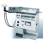
ENT diagnostic &
treatment units (ENT Exam Unit) |
Customised solutions for individual needs
The form and function of ENT Examination Unit workstations has
changed considerably over the past 50 years.
The BASIC PLUS line represents a temporary limit to
this long phase of development. All units are
designed to satisfy the highest possible
requirements.
Clearly organized and structured work station
The unit's modular design ensures clean lines and
has a reassuring effect on the patient.
Practical layout of functional elements
All the elements are positioned within easy reach of
the doctor and are also designed for single-handed
operation.
The instrument trays, preparation tray and waste
containers are situated close to the patient, whilst
the suspended cables of the light sources are
ideally placed in the centre of the unit for
protection. There are plenty of drawers and storage
compartments with space for consumable materials.
Flexibility when assembling the unit
There is almost no limit to the individual��s choice
of layout thanks to the unit's modular design and
the equipment available.
A variety of options
The BASIC PLUS line is built up around the ENT
diagnosis and treatment unit which is available in
three different widths. All versions may be used as
a ��stand-alone�� system or may be extended as desired
with additional modules (e.g. endoscopy centre,
instrument cabinet, desk attachment, plateau, etc.).
There is also the option of a left-handed version.
Basic model
��Compressed
air unit complete with fine regulation handpiece, 3
spray bottles, Politzer attachments, flow limiter
and pressure gauge
��Suction
unit with non-vibrating, smooth-running vacuum pump,
34 l /min, max. vacuum 93%
��Mirror
reheating unit
��Instrument
surface with Plexiglas cover
��Instrument
drawer
��Storage
drawer
��Open
compartment with electric socket for additional
equipment
Optional Extras
��38��C
warm water rinsing device with autoclavable water
filter unit
��Single,
double or quadruple cold light source, each 150 W
halogen
��Automatic
on/off switch and disinfection time control via
light barrier (or light switch control)
��Spittoon
on swivel arm with second suction hose
��Automatic
liquid container discharge
��Suction
tube cleaner with exchangable, autoclavable
stainless steel adapter
��Mirror
preheating unit for more than 50 mirrors of
different sizes
��Heated
instrument surface
��Instrument
cabinet
��Desk
module Type L, Type S
��High-plateau
with concealed sockets
��Swivel
support for additional equipment
��Integration
of practice's EDP system into the equipment concept
��Microscope
holder with electricity supply
��Stainless
steel edges to protect areas of particularly high
wear
��Melamin
drawers holding disinfectant solutions
��Built-in
waste bin for infectious wastes, syringes, etc.
��Pedal-operated
waste bin
��Mobile
on double castors
ORL3003
A complete ENT unit (ENT Exam Unit) that offers an unbeatable range
of rational solutions to specific problems combined
with the most sophisticated state of the art
technology. All functions are ergonomically
arranged.
Even with all accessories attached, compactness and
convenience are ensured.
Endoscopy quivers
The warming quivers and disinfection quivers can be
placed in the extension arm, so that an ergonomic
access to the scopes is guaranteed. If endoscopy
quivers are ordered in the extension, we recommend
to place the light source underneath the extension
beside the water filter. If not mentioned in the
order separately, cold light source will be
delivered on the frontside beside the melamin
drawer.
Suction pipe cleaner
A must for every ENT unit (ENT Exam Unit). Protects the suction tube
from clogging and disinfects the suction system
between the examinations. The cone shaped adapters
allow to clean the tubes and pipes of different
diameters. Due to their steel body and chrome-plated
surface, the adapters are easy to clean and to
autoclave. A couple of adapters will be delivered
with the unit for exchange.
Upon request the ORL 3003 L can be delivered with a
big ear rinsing funnel on a swivel arm. It is kidney
shaped and further detachable for cleaning.
Ear wax separator and noise reducer are further
details to increase the operating comfort.
Illuminated writing surface
Upon request the writing drawer/ dressing plate can
be delivered with illumination and automatic on /off
switch for illumination. Thus it doubles its
function as a writing surface (holder for patient
files and illumination of X-rays).
SWINGO
For clinics and private offices
Steel casing mobile on 4 castors
Large instrument surface with aluminium shelfs for
instrument compartments
Aluminium dust cover for instrument surface
Waste container with foot pedal
Container for used instruments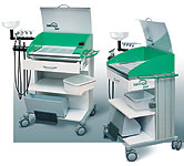
Support for additional equipment like HF-surgery
unit
Compressed air system, with 3 spray bottles and
politzer olives, 0.1�C 5 bar
Powerfull motor suction unit with 32 Ltr./min. and �C
0.95 bar max. vacuum
Warm water rinsing device 38��C with autoclavable
steel handle
Water filter system for rinsing device protects from
legionelles and pseudomonas. The filter is
autoclavable as well.
Kidney shaped ear rinsing funnel for the connection
to the motor suction tube
1 big storage drawer underneath the instrument
surface
Mirror heating unit single handed heating function
Cold light source 1 or 2 outlets with 150 watt
halogen lightpower
Smart
The mobile ENT Exam Unit SMART is built for the most
different areas of application.
It is usable for bed-side treatment, in operating
rooms, outpatient clinics and private offices.
Due to antistatic rubber castors, it is mobile and
can easily be adapted to any possible location.
The highly competitive suction unit is equipped with
comfort functions like automatic suction activation
and integrated suction pipe cleaner.
Up to three Halogen light sources are available for
endoscopic application.
2 x 150 W Halogen outlets with concave reflector
lamps, two layer thermal shield,
two intensity settings�� |
| Article Source: |
| http://www.rosslynmedical.com/en/r/product_catalogue/ent/ent_diagnostic_treatment_units/ |
|
Back To Top |
| �� |
|
ENT Doctors in New York
and New Jersey |
ENT and Allergy Associates (ENTA) is the largest
ENT doctor, allergy and audiology practice in the
tri-state area, with more than 30 offices in
Westchester, Putnam, Orange, Dutchess, Rockland, and
Nassau counties in New York, New York City
(NYC-Manhattan) and Bergen, Hudson, Middlesex,
Morris, Passaic and Somerset counties in New Jersey.
We offer the convenience of a large group practice,
with multiple office sites, evening and weekend
hours and prompt appointment scheduling. Our
practice features state-of-the-art care for ear,
nose, throat and allergy disorders, and offers a
variety of highly specialized procedures that
utilize the latest medical technology.
Allergy Doctors and Allergists in New York and New
Jersey
Our physicians (ENT doctors and allergists) and
licensed audiologists have the finest training and
experience and can treat a variety of conditions.
Our ENT doctor and allergist services include
diagnostics and treatment for sleep apnea and other
sleep disorders, sinusitis treatment, sinus surgery,
allergy treatments, facial plastic surgery, head and
neck surgery, laser surgery, nasal and laser
endoscopic sinus surgery, voice disorders, pediatric
ENT care, pediatric ENT Exam Unit, hearing disorders,
digital hearing aids, rhinoplasty, vertigo
treatment, and more.
ENT and Allergy Associates has a clinical alliance
with the Mount Sinai Medical Center for the
treatment of patients with head and neck disorders,
and has established a first-of-its kind program for
head and neck cancer screening. |
| Article Source: |
| http://www.entandallergy.com/ |
|
Back To Top |
| �� |
|
ENT doctors release
national guideline on treatment for common cause of
dizziness |
Published: Saturday, November 1, 2008 - 08:22 in
Health & Medicine
The American Academy of Otolaryngology �C Head and
Neck Surgery Foundation (AAO-HNSF) will issue a
comprehensive clinical guideline to help healthcare
practitioners identify and treat patients with
benign paroxysmal positional vertigo (BPPV), one of
the most common underlying conditions that cause
dizziness. The guideline emphasizes evidence-based
recommendations on managing BPPV, the most common
vestibular (inner ear) disorder in adults. BPPV is a
disorder that causes feelings of vertigo, dizziness,
and nausea. Episodes of BPPV can be brought on by
abrupt changes in movement, like standing up or
turning the head suddenly. The condition usually
begins to affect people after the age of 50, but it
can affect younger patients.
"Approximately 5.6 million medical appointments per
year in the United States can be attributed to
complaints of dizziness," said Neil Bhattacharyya,
MD, chair of the multidisciplinary BPPV Guideline
Panel. "We know now that anywhere from 17 to 42
percent of these patients will ultimately receive a
diagnosis of BPPV. Unfortunately, proper diagnosis
and treatment for those suffering is often delayed
due to a lack of standardized diagnostic steps and
relative unawareness of effective treatment
options."
The primary purposes of the new AAO-HNSF guideline,
for patients 18 years and older, are to improve
quality of care and outcomes for BPPV by improving
the accurate and efficient diagnosis of the
condition, reducing the inappropriate use of
suppressant medications, decreasing the
inappropriate use of ancillary tests such as
radiographic imaging and vestibular testing, and to
promote the use of effective repositioning maneuvers
for treatment.
Expenses relating to the diagnosis and treatment of
BPPV cost the U.S. healthcare system approximately
$2 billion per year. Additionally, 86 percent of
patients suffer some interrupted daily activities
and lost days at work because of BPPV.
Fortunately, BPPV can be readily diagnosed by
clinicians in an outpatient setting most of the time
without complicated testing. Once a proper diagnosis
has been made, simple, effective treatment options
are available to relieve symptoms quickly.
Some of the key recommendations
ENT Examination Unit of the guideline
include:
☺A
strong recommendation for clinicians to diagnose
posterior semicircular canal BPPV with an
office-based diagnostic test (the Dix-Hallpike
maneuver, detailed within the guideline).
☺A
recommendation for clinicians to also test patients
for a second type of BPPV affecting the lateral
semicircular canal when initial testing is not
conclusive (using the supine roll test).
☺Clinicians
should differentiate BPPV from other causes of
imbalance, dizziness, and vertigo.
☺Clinicians
should question patients with BPPV for factors that
modify management including impaired mobility or
balance, CNS disorders, a lack of home support, and
increased risk for falling. These recommendations
will help prevent some of the dangerous morbidities
from BPPV.
☺Clinicians
should not obtain radiographic imaging or vestibular
testing in a patient diagnosed with BPPV, unless the
diagnosis is uncertain or there are additional
symptoms or signs unrelated to BPPV that warrant
testing.
☺Clinicians
should not routinely treat BPPV with vestibular
suppressant medications such as antihistamines or
benzodiazepines.
☺For
patients who are initial treatment failures,
clinicians should evaluate them for persistent BPPV
or underlying peripheral vestibular or CNS
disorders.
☺Clinicians
should counsel patients regarding the impact of BPPV
on their safety, the potential for disease
recurrence, and the importance of follow-up.
The guideline was created by a multidisciplinary
panel of clinicians representing the fields of
otolaryngology, audiology, emergency medicine,
physical medicine and rehabilitation, geriatrics,
physical therapy, family physicians, neurology, and
chiropractics. |
| Article Source: |
| http://esciencenews.com/articles/2008/11/01/ent.doctors.release.national.guideline.treatment.common.cause.dizziness |
|
Back To Top |
|
|
�� |
�� |
|
|
|
| |
|