PK-6650
Colposcope digital imaging system
Introduction:
Combined with digital imaging technology and
computer, colposcope digital imaging system is the newest
gynecological examination equipment with clear image and
simple operation. It has replaced the traditional
examination mode. The minor pathological changes displayed
on the screen provide accurate evidences to the doctor for
the examination of erosion and cancer. The observed image
could be collected, frozen, zoomed in and saved with the
help of the advanced image and words process system. This
equipment supports remote consultation as an effective
workstation, adapting to the trend of digitization and
modernization of hospital.
Major function:
Dynamic video process
Display the input dynamic video; real-time of
freezing, observing, capturing, display, saving of the
image; display of image in full screen; adjusting of the
brightness, contrast, and saturation.
2. Static image process
Duplicating, zoomed in, rotating of the
chosen part; kinds of image display modes, such as
linearity, logarithm, exponent and balance; take note on any
spot at will; measure the diameter, girth and area of the
suspected pathological parts
Input, search and print the inspection
report.
Extended-definition scene made by Sony,
Japan, 560,000 pixels; 220 times zooming; cold light source
compensation
Diagnosis scope: Cervical erosion, cervical
polyp, swollen gland, leukoplakia vulvae and venereal wart。
(Laptop
computer) Portable Digital electronic vaginoscope (Contain
software)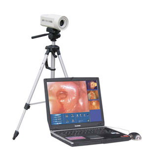
Characteristics of the working station system ;
Import SONY 1/4 high-resolution-factor digital color CCD ;
470,000 dot image elements, horizontal resolution factor
above 480 lines ;
Image stepless magnification of 1 to 30 times ;
Video and S-video mode output ;
High-speed automatic gathering or manual gathering control ;
Automatic self-balance adjustment and electronic numerical
reading technology ;
Automatic adjustment of self-balance ;
Focusing distance 10 to 800mm ;
Unique design of light source;
Adopting super-low ring multipoint white cold-light source ;
Even brightness bright hue, a small size and a long life
length ;
Software character :
1.Powerful computer imaging, data management and disease
analysis . It has WINDOWS98 Chinese operating system with
the functions of image collecting, displaying, processing
and report printing and storage.
2. Control the focus of CCD, the scale of the easy real-time
picture through the computer , help the doctor choose the
clearer , more high-quality picture greatly
3. The special optical filtering system, can show the false
color of many kinds of dynamic filters such as being red,
green, blue in real time.
4. Abundant case atlas base, providing a large number of
case atlases. It is very convenient to make comparative
analysis of atlas, which is helpful for patient's diagnosis.
Produce the content of reporting automatically.
5. The beautiful print report, an abundant diagnosis
template,
6.large store, case history and the information of checking
are store at the same time, the picture is more than
100,000.
7. 17inch high-resolution-factor color large screen display,
adopt HP ink high-resolution-factor color image output.
Portable
Digital electronic vaginoscope (Contain software)
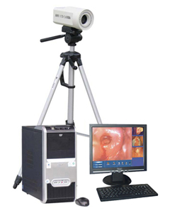
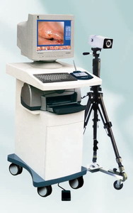
System configuration:
Digital vaginoscope lens
Advanced computer configuration
17’ flat color monitor with high resolution
Triangle stand
Easy to assemble and move, high quality and reliable
performance
Optional:
LCD monitor
Aluminum alloy packing case
Color jet printer
Software character :
1.Powerful computer imaging, data management and disease
analysis . It has WINDOWS98 Chinese operating system with
the functions of image collecting, displaying, processing
and report printing and storage.
2. Control the focus of CCD, the scale of the easy real-time
picture through the computer , help the doctor choose the
clearer , more high-quality picture greatly
3. The special optical filtering system, can show the false
color of many kinds of dynamic filters such as being red,
green, blue in real time.
4. Abundant case atlas base, providing a large number of
case atlases. It is very convenient to make comparative
analysis of atlas, which is helpful for patient's diagnosis.
Produce the content of reporting automatically.
5. The beautiful print report, an abundant diagnosis
template,
6.large store, case history and the information of checking
are store at the same time, the picture is more than
100,000.
7. 17inch high-resolution-factor color large screen display,
adopt HP ink high-resolution-factor color image output.
8.Fast and convenient data inquiry. Supporting Internet
|