|
ABSTRACT
The use of intraoperative navigation
systems in neurosurgery has
increased rapidly. The Neuronavigation Microscope(Neurosurgery
(Neural,neurosurgical) Operation (surgery,surgical,operating)
microscope,Neurosurgery (Neural,neurosurgical)
microscope)
Integration (NMI) system consists of
a microscope (Zeiss, Germany)
combined with the StealthStation (Medotronic,
U.S.A.) including light emitting
diodes, a dynamic reference frame
with light emitting diodes, an
optical digitizer with camera array
and a computer workstation. The aim
of this study was to determine the
usefulness of the NMI system for
neurooperating operations. Between
April 2003and March 2011, the
authors used the NMI system in 367
patients undergoing neurosurgical
operations at Kagawa University
Hospital. Because the navigational informations could be superimposed
onto the microscope view, accurate
locations of tumor and normal
anatomical structures could be
obtained before
skin incision. During the
operations, the surgeons did not
need to turn away from the surgical
field or to use a bulky pointer.
Catheter applications along the
tumor borderline guided by the NMI
system could be useful for glioma
Operation. Deep seated lesions
including intraventricular or
intra-axial tumors couldbe removed
through accurate and minimal
corticotomy. For transsphenoidal
Operation, pituitary tumors could be
safely removed without X-ray
imaging. For the skull base
Operation,
the navigational information was not
affected by the brain shift during
the operations. The registration
assessment deviations were within 2
mm and the real anatomical
deviations were within 3 mm. The
authors’ findings suggest that the NMI system can provide valuable and
reliable intraoperative navigational
informations during neurosurgical
operations.
INTRODUCTION
The use of intraoperative navigation
system in neurosurgery (Neurosurgery
(Neural,neurosurgical) Operation (surgery,surgical,operating)
microscope,Neurosurgery (Neural,neurosurgical)
microscope) has increased
rapidly (Brinker,1998: Enchev, 2009:
Germano, 1999: Golfinos,1995:
Roessler, 1997: Samii, 2000: Sipos,
1996:Takizawa, 1993: Uluc, 2009:
Watanabe, 1991:Yoshikawa, 2006).
Computer-assisted neuronavigationfor
the intraoperative viewing of
anatomical landmarks is increasingly
used for the operating removal of
intracranial lesions. Neuronavigation simultaneously
represents a
complex,multimodal,information-based,
highly adaptable technique,method,
or device using frameless stereotaxy
for precise intraoperative guidance,
orientation, and localization, with
consequently greater operating
precision and possibilities for
preoperative virtual simulation and
postoperative analysis of the
operating procedures (Enchev, 2009).
The Neuronavigation Microscope(Neurosurgery
(Neural,neurosurgical) Operation (surgery,surgical,operating)
microscope,Neurosurgery (Neural,neurosurgical)
microscope)
Integration (NMI) system is composed
of a microscope (Zeiss, Germany) and
the StealthStation® (Medtronic,
U.S.A.) that includes light emitting
diodes, a dynamic reference frame
with light emitting diodes, an
optical digitizer with a camera
array and a computer workstation.
The aim of the study was to evaluate
the suitability and usefulness of
the NMI system for neurosurgical
operations(Neurosurgery
(Neural,neurosurgical) Operation (surgery,surgical,operating)
microscope,Neurosurgery (Neural,neurosurgical)
microscope).
BACKGROUND
Operating
microscope(Neurosurgery (Neural,neurosurgical)
microscope) have improved
tremendously since they first
entered the operating room. Today
they offer good magnification
without significant aberrations,
sufficient illumination without
excessive heat, and satisfying
stability without sacrificing
operational flexibility. Modern
microscopes(Neurosurgery (Neural,neurosurgical)
microscope) have already started to
combine multiple visualization
techniques (Uluc, 2009).These
microscopes can display
intraoperative navigation
informations simultaneously.In
contrast, neuronavigation
simultaneously represents a complex,
multimodal,informationbased
informationbased,widely adaptable
technique, method, or device using
frameless stereotaxy for precise
intraoperative guidance,
orientation, and localization, with
consequently greater operating
precision as well as the potential
for preoperative virtual simulation
and postoperative analysis of the
operating procedure.
MAIN FOCUS OF THE CHAPTER
Registration of the Neuronavigation
Microscope(Neurosurgery
(Neural,neurosurgical) Operation (surgery,surgical,operating)
microscope,Neurosurgery (Neural,neurosurgical)
microscope) Integration (NMI) System
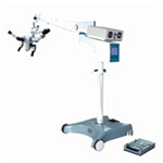
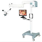
Intraoperative image guidance was
achieved using a NMI system composed
of a microscope
(Zeiss, Germany) and StealthStation®
(Medtronic,U.S.A.) (Figure 1).
Accuracy in image-guided surgery
directly depends on how well the
radiographic images and surgical
anatomy of the patient is
correlated. The process of
establishing a computer map between
all locations on the images and the
corresponding anatomical locations
in the operating field is called registration.Registration is
initiated using the disposable skin
fiducial markers (Medtronic, U.S.A.)
and surface landmarks, which
correlate with the same points on
the three-dimensional CT/MRI surface
reconstruction of the skin. The
StealthStation system (Medtronic,
U.S.A.) employs two different
methods for achieving object
alignment and transformation:
PointMerge™ registration and
SurfaceMerge™ registration.
PointMerge registration is the
primary method and must be used for
each Operation. SurfaceMerge
registration is a secondary method
employed in the spinal application
as a refinement to the PointMerge
method. Both methods attempt to find
the best coordinate system
transformation between image space
and operating space by aligning the
user-defined objects contained
within them. The difference between
the
methods lies in how the computer
represents the objects internally
and how the computation of the
‘best’ alignment changes with this
representation.
www.irma-international.org/viewtitle/70855/
|