|
[Abstract] Objective
through our department since March
2009 - July 2010 using
dental
operating microscope (DOM) on the
treatment of refractory pulp periapical lesions cases, evaluation
of
dental surgery microscope
refractory root canal treatment The
clinical application of results.
Transferred to our hospital and
outside the hospital ordinary endodontics clinic to 89 patients, a
total of 106 root canal refractory
cases, including root canal
calcified root canal filling plug,
instrument fracture, omissions root
canal mandibular first second molar
"C"-shaped root canal, surgical
microscope with ultrasonic root
canal treatment instrument and other
microsurgical instruments, foreign
body removal, root canal preparation
processing. Results The total
effective rate of 86%. Conclusion:
The
dental operating microscope
(Dental (dentistry) Operating (operation,surgical,surgery)
microscope) with the
relevant equipment can make more
refractory teeth root canal
treatment, so as to preserve the
tooth.
Key words surgical microscope(dentistry Operation microscope,Dental
(dentistry) Operating (operation,surgical,surgery)
microscope) root
canal treatment ultrasonic
instruments
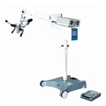
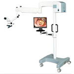
Dental operating microscope (DOM,
dentistry Operation microscope,Dental
(dentistry) Operating (operation,surgery)
microscope) amplification
effect and good lighting advantage
due to its traditional endodontic
treatment process from blindness to
visual, handling a variety of causes
root canal blockage, instrument
fracture root canal calcification,
"C" shaped canal, and achieved good
results. This article by our
hospital DOM treatment of refractory
teeth Sui cases of periapical
lesions, to evaluate the clinical
effect.
1 Materials and methods
1.1 Case Source
Our department since March 2009
-2010 in July, a total of 89 cases
of admissions to our hospital and
outside the hospital GOP referral
refractory teeth Sui periapical
lesions cases, a total of 106 root
canal.
1.2 Devices
Root canal surgery microscope
(dentistry Operation microscope,Dental
(dentistry) Operating (operation,surgical,surgery)
microscope); ultrasonic root canal
treatment, ultrasonic working tip;
microscopic reaming needle; probing
teeth Sui; microscope dedicated
surface reflection mouth mirror root
canal surgery; rubber dam;: 8 #, 10
# K filing, EDTA paste.
1.3 Methods
Observe whether the symptoms of
dental (dentistry Operation
microscope,Dental (dentistry)
Operating (operation,surgical,surgery)
microscope)fear before using dental
microscope surgery(Dental
(dentistry) Operating (operation,surgical,surgery)
microscope), the
establishment of the medical records
of all cases operating under rubber
dam. Before the beginning of the
operation, carefully read the
preoperative X-ray film to
understand the root canal
morphology, root canal wall
thickness, root canal blockage,
break location and length of the
instrument. Looking for a root canal
mouth, or open up the calcified root
canal: adjust the light source,
Dental (dentistry) Operating (operation,surgical,surgery)
microscope, the use of
ultrasound tip to remove calcified
dentin revealed root canal port,
with the microscopic reaming needle
and EDTA paste gradually dilate root
canal removal The broken
instruments: Dental (dentistry)
Operating (operation,surgical,surgery)
microscope,
ultrasonic tip to remove broken
instruments around the root canal
wall dentin, it gradually loosened
up and pop-under ultrasonic
vibration. Removal of zinc phosphate
water door D, the plasticizing
liquid, gutta percha: under a
microscope, using appropriate
ultrasonic tip vibration broken root
canal
Of obstruction, to get through the
root canal. Mandibular second molars
"C" shaped canal preparation: under
a microscope using ultrasonic
instruments with hand or machine
endodontic instruments, preparation
of the root canal and
Isthmus.
1.4 Data Retention
The use of digital image acquisition
card storage on the computer, or use
the built-in lens optical camera,
each of the cases were taken before
surgery, postoperative X-ray.
2 Results
The dental surgical microscope
(dentistry surgery microscope,Dental
(dentistry) Operating (operation,surgical,surgery)
microscope)clinical application results
The statistics overall success rate
of 86%.
3 Discussion
Through the DOM so difficult root
canal treatment in our hospital
evaluation, success rate of 86%.
Instrument fracture in the root
canal root canal preparation was
2.02% -2.61% [1], often cause
patient disputes. Removal of broken
instruments surgery to enhance
communication. We remove the broken
needle is mostly located in the root
in 1/3, and also individual located
in apical 1/3. However, fewer cases,
a high success rate, instrument
fracture location is significant
factors that affect the success rate
of treatment and duration of
treatment, whether in the Dental
(dentistry) Operating (operation,surgical,surgery)
microscope (dentistry surgery
microscope,dentistry surgical
microscope,
dentistry operating microscope)fully exposed broken
instruments, the operator cleaning
observe the entire
The course of treatment is a
prerequisite for microscopic root
canal treatment success. Principle,
as long as it is fully exposed to
the full length of the broken
instruments, 1/3 will be able to
remove the broken material [2] root
canal calcification is more common
in clinical practice, mainly for
small developing unclear X-existing
on-chip root canal or root canal; 30
Dental (dentistry) Operating (operation,surgical,surgery)
microscope(dentistry surgery
microscope,dentistry surgical
microscope,
dentistry operating microscope), using ultrasound
work sharp removal the calcified
dentin lighter in color, by means of EDTA paste root calcification
microscopic root canal treatment,
found most of the root canal
calcification in the apical 1/3, 8
#, 10 # K filing, can dredge the
root canal. In the visual case, a
more accurate judgment of the
cutting portion of the dentin, and
effectively avoid the occurrence of
the offset and the root of the root
canal wall perforation. Years for
the treatment of dry Sui, to be
crowns, to do root canal treatment,
root canal calcification, mostly
molars unshaded apical root canal
curvature, calcification
heterogeneity (dental operating
microscope) Dental
(dentistry) Operating (operation,surgical,surgery)
microscope, it is difficult to
distinguish shades of color
ultrasound to get through to the
apical 1/3, for each direction is
difficult to grasp, not to continue
treatment to avoid root canal side
wear using the DOM in the treatment
of a variety of causes root canal
clogged removal of root canal
fillings and broken instruments in
the evaluation a success rate of
100%, the root canal filling gutta
percha, zinc phosphate or
plasticizing blockages in the DOM
visualization circumstances, using
ultrasound tip blind to get through,
causing the risk of root canal side
wear DOM looking for a unique effect
of missing root canal alignment clog
its vibration broken, you can expand
through root canal [3], to avoid the
traditional treatment. Omissions
root canal on a segment of the root
canal calcified root canal mouth
ectopic or too small medullary
cavity entrance. Occurs most
frequently missing mesiobuccal root
of maxillary molar root canal (mb2)
or root canal (mb3), second or third
root canal maxillary premolar,
mandibular
The molars third mesial root canal
as well as the second or third
distal root canal [4]. Root fusion
in conical or square cross-section
on the root canal was the "C"-shaped
root canal system known as the "C"
shaped canal system commonly used in
clinical buccolingual X-ray
diagnosis is difficult using
conventional root canal treatment
technology preparatory root canal
may occur when part of the root
canal wall is over-preparation,
while the other Ministry
Failure to clean up the phenomenon
of sub root canal wall. The form of
the root canal orifices clear vision
microscopic root canal treatment
technology for enhanced light source
root canal preparation and
amplification conditions under avoid
excessive
Tooth preparation, when the upper
part of the root canal mouth open,
light deep into the root canal, and
easy to find residual necrotic
tissue and dentin debris, to
facilitate the identification of the
site and the effect of the root
canal.
Dental operating microscope (Dental
(dentistry) Operating (operation,surgical,surgery)
microscope) with the relevant
equipment can make more refractory
tooth root canal treatment, so as to
preserve the tooth.
References
[1] XIA Ning, Zhao Xinchen, high
Xiaoyan, root canal treatment, 75
cases of the instrument fracture of
clinical analysis [J] Oral Medicine.
Aspect, 1999.15 (2
) 103 ~ 105.
[2] Ling Jun-qi, Xi Wei. Microscopic
root canal treatment efficacy and
influencing factors [J] of Shanghai
Oral Medicine, 2006,15 (1): 1-6.
[3] Ling Jun-qi, Xi Wei, Gao Yan,
the application root canal
microscope and ultrasonic
instruments processing blocking the
evaluation of the efficacy of root
canal [J] Journal of Oral Medicine
Journal, 2003,38 (5): 9-11.
[4] Fan Bing. Microscopic root canal
treatment dental(Dental (dentistry)
Operating (operation,surgical,surgery) microscope)(dentistry surgery
microscope,dentistry surgical
microscope,
dentistry operating microscope) endodontics. 2
Beijing: People's Health Publishing
House, 2003:270 ~ 276 Keywords
: Surgical microscope root canal
treatment ultrasonic instruments |