|
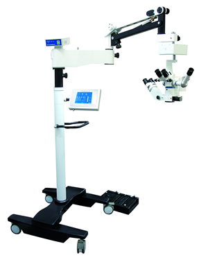
Model LZL-16 Eye surgery microscope
Ophthalmic
surgical microscope,Ophthalmic
operation microscope,Ophthalmic
surgery microscope,Ophthalmic
Operating microscope
Ophthalmic-specific:
1.High-definition continuous
variable magnification
optical system
2. coaxial illumination of
cold light source dual bulb
conversion
3. foot control electric
focusing and foot control
lighting switch
4. new balance rack mover
with X-Y plane
5.Microcomputer centralized
control system and the image
system
6. Optional splitter, the
Teach microscope and digital
camera system and CCD camera
system as well as the xenon
lamp lighting
Technical parameters:
Microscope
Magnification of eyepiece:
10/22B
Working Distance: 200mm
The primary mirror
Magnification: 4-24 electric
stepless zoom
Secondary mirror
magnification: 6, 1,16
Field of view diameter:
58mm-11mm
IPD adjustment range:
55mm-75mm (continuously
adjustable)
Diopter adjustment range: ±
6D (continuously adjustable)
The speed and range of fine
focusing: ≤ 2mm / s, 40mm
X / Y moving speed and
range: ≤ 2mm / s, 40mm *
40mm
Light Source
Lighting type: 6 ° +0 °
coaxial illumination
Lighting bulbs: 15V/150W
halogen lamps (optional 250W
xenon lamp)
Lighting control: continuous
digital regulation
Maximum illumination of the
object plane: ≥
80000-120000Lx
Optional xenon lamp
lighting, illumination of up
to 180,000 Lx
Membrane touch switch and
LCD display design, reliable
Maintain the usual
parameters of the 12 doctors
Frame
Arm stretch radius: 1200mm
Vertical adjustment range:
450mm
Uses:
LZL-16-type surgical
microscope carefully
designed by the famous
optical experts machine
adopts advanced modular,
digital, personalized design
concepts, products widely
used in ophthalmic
microsurgery.
Features:
1.The main microscope using
a state-of-the-art parallel
light electric continuous
zoom technology, zoom factor
of 0.45-2.4,
high-definition, large depth
of field, layering, can meet
a variety of deep surgical
observation; eyepiece
operative field, the
operation The distance is
less than 40mm, operative
prolonged eye, the body will
not fatigue.
2.Instrument uses coaxial
cold light source fiber
optic lighting, excellent
three-dimensional, with
retinal protection devices
and foot control lighting
switch (to facilitate
vitrectomy).
3.Vice microscope for three
drum zoom, and the main
microscope at a 90 °
postural, the same optical
path.
4. independent electrical
control system, installation
and maintenance convenient.
5. Tune pour institutions
the surgery face special
viewing angle can be set.
6. microcomputer program
control, stepless dimming,
speed control system.
7.With foot controlled XY
plane mover, moving range XY:>
40mm. Can be reset to zero.
Optional Accessories:
Splitter, monocular Teach
mirror, digital camera
interface, 1/3 inch CCD
camera interface, expert
video image processing
system, and the optional
xenon lamp lighting. |