|
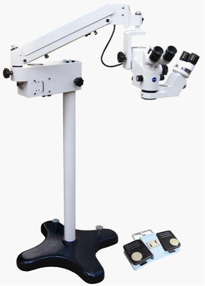
Model LZJ-5D ophthalmic surgical microscope
Eye surgical
microscope,Eye
operation microscope,Eye
surgery microscope,Eye
Operating microscope
use
Model LZJ-5D ophthalmic
surgical microscope, can be
applied to microscopic
surgery.
Second, characteristics and
main performance index
(a) characteristics :
1 and the Lord microscope
using parallel light
electric five file variable
times technology, binoculars
barrel to 45 °. Instrument
imaging is clear, the depth
of field is big, a wide
field of vision, working
distance for 200 mm.
2, binocular assistant
mirror and the main
microscope into 90 DHS
position.
3, instrument using coaxial
cold light source inside
lighting system, intensity
of illumination was 50000
lx.
4, instrument optional
monochrometer interface, can
match monocular teach
mirror, photographic
interface, CCD camera
interface.
(2) the main performance
index
1,
(Eye
surgical microscope,Eye
operation microscope,Eye
surgery microscope,Eye
Operating microscope)primary mirror
|
Handwheel and value
|
0.4 |
0.6 |
1 |
1.6 |
2.5 |
|
magnification |
3.5 |
5.4 |
8.5 |
13.5 |
21 |
| Field
of view(mm) |
68 |
43 |
27.5 |
17.5 |
11 |
2, (Eye
surgical microscope,Eye
operation microscope,Eye
surgery microscope,Eye
Operating microscope)assistant mirror
|
magnification(X) |
6x |
|
Field of view(mm) |
35.3 |
* the Lord, deputy
microscope eye distance
regulating range 55 ~ 75 mm
* trimming focal range 35 mm
* cross arm maximum
extension length of 1080 mm
* frame vertical lift range
450 mm
* lighting spot diameter Φ
65 mm
* microscope before and
after the pitch range not
less than ± 30 °
3,
(Eye surgical microscope,Eye operation microscope,Eye surgery microscope,Eye Operating microscope)
optical parameters
3.1 optical system between
the magnification difference
is not more than 1.5%.
3.2 optical system between
the field consistency:
things mirror in-plane
vertical migration quantity
is not more than 0.2 mm,
horizontal offset no greater
than 0.4 mm.
3.3 or so between the
optical system of the focal
length difference is not
more than 5 mm.
3.4 the highest times field
resolution not less than 63
line/mm.
3.5 or so between the field
like inclined not greater
than 2 °.
3.6 optical system exit
pupil height difference set
visibility moment value in
the 0 bit not more than 1.5
mm.
3.7 distance minimum
adjusting range 55-75 mm.
3.8 visibility minimum
adjusting range + 5 - - 5 m
- 1.
3.9 field relative tolerance
is not more than 2.5%.
3.10 object surface spot
center illumination not less
than 15000 lx.
4,
(Eye surgical microscope,Eye operation microscope,Eye surgery microscope,Eye Operating microscope)mechanical properties
Micro lift range not less
than 35 mm.
Each activity joint
operation should be smooth
and flexible, accurate
positioning, Lock the stable
and reliable, and can not
have jitter, automatic sink
or card hysteresis. Frame in
any position should be in
good balance.
5,
(Eye surgical microscope,Eye operation microscope,Eye surgery microscope,Eye Operating microscope)electrical properties
Safety category: I kind of
equipment. Single-phase
network power supply: 220 v,
50 hz.
Input power 220 va, fuse
capacity of 1.5 A x 2, A
multiply one.
5.1 surgery microscope work
when the noise is not more
than 65 db.
5.2 protective grounding
impedance of not more than
0.2 Ω.
5.3 normal operating
temperature of the earth
leakage of not more than 0.5
mA, a single fault condition
should be not more than 1 mA
5.4 normal working
temperature dielectric
strength:
A) network power section and
has protective earthing
shell parts should be able
to withstand between 50 hz
and 1500 v sine wave test
voltage 1 min, no breakdown
and flashover phenomenon (a
- al);
B) intermediate circuit
(transformer secondary 21 v
winding and shell between)
should be able to withstand
50 hz and 500 v sine wave
test voltage 1 min, no
breakdown and flashover
phenomenon (A - al).
5.5 foot switch used in the
operating room with
prevention into liquid
requirements.
5.6 the technical
description and
specification merger. Users
to repair if need relevant
technical data, can all
alone to manufacturer line
take.
5.7 this equipment no
electromagnetic
compatibility special
requirements, portable and
mobile RF communication
equipment had no effect on
this equipment. This
equipment without source and
electrostatic put power
supply, this system for
attachment within the
increase and change will not
lead to the increase of the
launching equipment and
immunity to reduce. The
equipment should not and
other equipment close or
layers use, if you must
close or layers use, should
be observed verification in
the use of the configuration
can be normal operation.
Three, installation
1, open the packing
container, check whether
conform to physical and
packing list.
2, first will stand for
good, will microscope lens
(the Lord, deputy
microscope) take out, it
will be on the left a shaft
insertion bending plate of
the bottom of hole, and the handwheel binding, again
will screw admiral shaft and
bending plate binding, and
the last in the after
electrify check each
function is normal use, such
as no problems can be pushed
into the operating room to
use.
Four, method of use
(1), in the surgery
microscope after the
installation, push them to
the right position surgery,
on the base of the two
brake, lock instrument, in
case microscope in surgery
about. If you want to
loosen, please tread down
locking lever next to the
pedal, the locking lever
will advance pop-up, thus
loosen instrument, this
instrument can move freely.
(2), switch on the power,
open frame on the power
switch.
(3), loosen the frame of all
a tight the handwheel (see
chart 1), mobile lens to
surgery surface (observation
surface) above (or front)
work focal length place (as
the lens focal length
decide) until the eyepiece
can see the observation
surface so far, lock on the
frame of the handwheel.
(4) and adjust the master
with the hand lens of the
eye from the size (figure 1,
8), to suit your distance.
(5), regulating drum become
times the handwheel (figure
1, 6), to the over-error of
2.5 (the largest
magnification).
(6), by foot pedal control
lens micro lifting device,
until see things surface.
(7), may be around eyes
vision not consistent, the
eyepiece of about objective
image definition is differ,
right now but screwing in or
spin out side of the
eyepiece.
Note: the left ocular
diopter position and
distance size on everyone
basic is fixed, adjusted the
is not allowed to
adjustment, such as surgery
surface does not clear
phenomenon, can adjust
working distance. (FIG. 5,
13)
(8), 6 x assistant mirror:
binocular assistant mirror
and primary mirror into 90
DHS position, according to
need to turn to the right or
left of the primary mirror,
as long as assistant mirror
turn to the left or right
position, and the main
mirror into 90 DHS postures,
adjust good visibility and
distance were observed.
(9), in the surgeon clear
observation, assistant
mirror observer can't see
clearly like, and the
surgeon is different, can
adjust the ocular diopter,
until clear so far. In the
process of operation
assistant mirror users
without the consent of the
the surgeon is can't
literally adjust the working
distance.
(11), the choose and buy a
(teach mirror, camera,
camera device) use.
(1), loosen 45 º eyepiece
group at the bottom of a
screw, take off the eyepiece
group.
(2), optical splitter
inserted his lens, location
is good, tighten the
monochrometer a screw.
(3), will eyepiece group
insert optical splitter,
location is good, tighten
the eyepiece group a screw.
(4), will teach mirror
insert monochrometer
interface (or so all can),
optical splitter another
side interface insert camera
or camera interface, would
connect the handwheel
tightening, and then put the
camera or CCD camera.
(5), and CCD camera with
signal wire and monitor and
video equipment connection,
can be realized in the
process of operation
SheLuXiang.
(12), after using it, you
should turn off light
switch, then turn off the
power switch.
(13), in lamp: loosen
bearing arm side department
of the four screws (figure
1, 12), upward force pull
out, can change the light
bulbs, in good again after
the original direction
insert, screw on screw can.
Five, maintenance and
matters need attention
(a) maintenance
1, use and storage
instrument place (generally
refers to the operating
room), should be kept clean,
pay attention to dust,
moistureproof, acid and
alkali resistant, etc have
corrosion volatile matter
qualitative erosion. Use
after application dust
shroud live.
2, special attention should
be paid to keep the optical
lens clean, fingers are not
allowed to touch the optical
lens surface. If the lens
surface dust, usable and
clean brush or brush wiped
the, also can use a
handkerchief to wipe gently.
If not careful hand met or
oil pollution, usable cotton
wool with a small amount of
ethyl ether ethanol mixture
(ethanol 30% ethyl ether
70%) is brushed try, wipe
try don't let the mixture
into the lens inside. In
order to avoid lens
degumming or mildew.
3, optical fiber is made of
many glass composition,
unfavorable had large
bending force, need bending,
curve radian but small (90 °
above), in order to avoid
broken, influence through
light, deposit should bend
coil.
(2) the matters needing
attention
1, this instrument is Ⅰ
class B equipment, request
supply network power supply
(220 v ± 22 v, 50 hz ± 1 hz)
has a good grounding line,
and when using please be
checked, lest cause security
risk. Instrument maximum
input power 220 va.
2, when replacing the fuse
please close instrument
power switch, unplug the
power plug, change the
specifications of the fuse
tube, in case produce
security risk.
3, in the operation such as
a bulb is damaged, it should
be timely enable spare lamp
lighting, change bulb must
be in stop 30 minutes after
tight, in order to avoid
high temperature scald.
4, instrument on the various
handwheel when use must be
binding, in order to avoid
accident. On the frame
regulating balance force
size the handwheel at the
balance arm after adjustment
is not active.
5, the instrument attached
electric schematic diagram
for reference only
maintenance, are subject to
change without prior notice.
6,
(Eye
surgical microscope,Eye
operation microscope,Eye
surgery microscope,Eye
Operating microscope)instruments and normal
working condition
A) environment temperature 5
℃ ~ 35 ℃;
B) relative humidity 30% ~
80%;
C) the atmospheric pressure
700 hpa ~ 1060 hpa.
Six, and the spare
accessories and choose and
buy a see packing list
Seven, instruments used
graphics, symbols,
abbreviations, etc
explanation
"Ι" said power supply switch
on,
"О" said power switch close.
"And a" said fuse to 2 amp;
 Said
brightness increase; Said
brightness increase;
 Said
brightness becomes Said
brightness becomes
 Said
microscope rise; Said
microscope rise;
 Said
microscope decline. Said
microscope decline.
Eight, the electrical
principle diagram
Nine, transportation and
storage condition: :
A) environment temperature
range - 40 ℃ ~ 55 ℃
B) relative humidity of 95%
or less
C) the atmospheric pressure
range is 500 hpa ~ 1060 hpa.
D) transport requirements
stipulated in the contract
according to the order.
Packaging good surgery
microscope should be stored
in a relative humidity no
more than 80%, no corrosive
gas and other harmful
substances, well ventilated
indoor. |