| Lumbar Discectomy
(hand
surgical microscope, orthopaedics ,orthopaedy, orthopedic,operating
microscope)
Introduction
Lumbar discectomy is a surgical procedure to remove part of a
problem disc in the low back. The discs are the pads that
separate the vertebrae. This procedure is commonly
(hand
surgical microscope, orthopaedics ,orthopaedy, orthopedic,operating
microscope)
used when a
herniated, or ruptured, disc in the low back is putting pressure
on a nerve root.
This guide will help you understand
•what surgeons hope to achieve
•what to expect as you recover
Anatomy
What parts of the spine and low back are involved?
Surgeons (Hand
surgical microscope, Orthopaedics, Orthopaedy, Orthopedic,
Operating
microscope)
perform lumbar discectomy chirurgery through an incision
in the low back. This area is known as the posterior region of
the low back. The main structure involved is the intervertebral
disc, which acts as a cushion between each pair of vertebrae.
The two main parts of the disc are the annulus and the nucleus.
Lamina bone forms the protective covering over the back of the
spinal cord. During
(hand
surgical microscope, orthopaedics , orthopaedy, orthopedic,operating
microscope)
, this section of bone is removed
over the problem disc. The surgeon also checks the spinal nerves
where they travel from the spinal canal through the neural
foramina. The neural foramina are small openings
(hand surgical microscope, orthopaedics ,orthopaedy,
orthopedic,operating
microscope)
on each side of
the vertebra. Nerves that leave the spine go through the
foramina, one on the left and one on the right.
Related Document: A Patient's Guide to Lumbar Spine Anatomy
Rationale
What do surgeons hope to achieve?
Lumbar discectomy can alleviate symptoms from a herniated disc
in the low back. The main goal of discectomy
(hand
surgical microscope, orthopaedics ,orthopaedy, orthopedic,
operating
microscope)
is to
remove the part of the disc that is putting pressure on a spinal
nerve root. Taking out the injured portion of the disc also
reduces chances that the disc will herniate again.These goals can be achieved using a traditional procedure, called laminotomy and discectomy
(orthopaedics ,orthopaedy, orthopedic,operating
microscope, hand surgical microscope), or with a newer method called
microdiscectomy
(orthopaedics ,orthopaedy, orthopedic,operating
microscope,hand surgical microscope). The traditional method requires a larger
incision and tends to require a longer time to heal.
Microdiscectomy is becoming the standard chirurgery
(orthopaedics ,orthopaedy, orthopedic, operating
microscope,hand surgical microscope)for lumbar disc herniation.Since the surgeon performs the with a
surgical microscope, he or she needs to make only a very small
incision in the low back. Categorized as minimally invasive
chirurgery, this OPS is thought to be less taxing on patients.
Advocates also believe that this type of OPS
(Hand surgical microscope,
Orthopaedics ,Orthopaedy, Orthopedic,
Operating
microscope) is easier to
perform, prevents scarring around the nerves and joints, and
helps patients recover more quickly.
Related Document: A Patient's Guide to Lumbar Disc Herniation
Preparations
How will I prepare for ops?
The decision to proceed with OPS
(Hand surgical microscope,
Orthopaedics ,Orthopaedy, Orthopedic,
Operating
microscope) must be made jointly by you
and your surgeon. You should understand as much about the
procedure as possible. If you have concerns or questions, you
should talk to your surgeon.
Once you decide on OPS, your surgeon may suggest a complete
physical examination by your regular
(Hand surgical microscope,
Orthopaedics ,Orthopaedy, Orthopedic,
Operating
microscope) doctor. This exam helps
ensure that you are in the best possible condition to undergo
the OPS.
On the day of your operating
(orthopaedics , orthopaedy, orthopedic, operating
microscope, hand surgical microscope), you will probably be admitted to the
hospital early in the morning. You shouldn't eat or drink
anything after midnight the night before.
Microdiscectomy
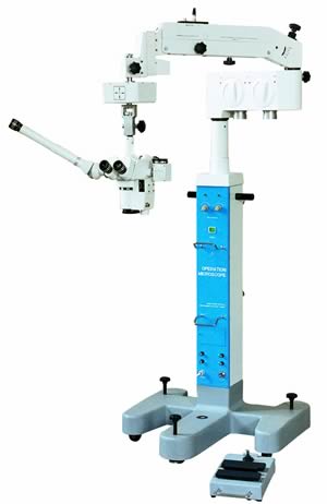
Surgeon performs microdiscectomy using a surgical
microscope (orthopaedics, orthopaedy, orthopedic, operating
microscope,hand surgical microscope,hand surgical microscope). A two-inch incision is made in the low back directly
over the problem disc. The skin and soft tissues are separated
to expose the bones along the back of the spine. An X-ray of the
low back is taken to ensure the surgeon works on the right disc.
A retractor (Orthopaedics,
Orthopaedy, Orthopedic,
Operating
microscope, Hand surgical microscope,Hand surgical microscope) is used to spread apart the lamina bones above and
below the disc. Then the surgeon makes a tiny slit in the ligamentum flavum, exposing the spinal nerves. A special hook is
placed under the spinal nerve root. The hook is used to lift the
nerve root, so the surgeon can see the injured disc.
Next, the annulus (outer ring) of the disc is sliced open.
Material from inside the disc is scooped
(Orthopaedics,
Orthopaedy, Orthopedic,
Operating
microscope, Hand surgical microscope,Hand surgical microscope)
out to ensure the disc
doesn't herniate again.Since only the injured portion is
removed, the disc is left intact and functioning. Then the
surgeon inspects the area around the nerve root and removes any
loose disc fragments. Finally, the nerve root is gently wiggled
to make sure it is free to move. If it can't move, the surgeon
also cleans around the neural foramen, the nerve passage between
the two vertebrae.
(orthopaedics, orthopaedy,orthopedic,operating
microscope, hand surgical microscope) When the nerve moves freely, the muscles and
soft tissues are put back in place, and the skin is stitched
together.
Infection
Infection following spine operating(orthopaedics,orthopaedy,orthopedic,operating
microscope, hand surgical microscope) is rare but can be a very
serious complication. Some infections may show up early, even
before you leave the hospital. Infections on the skin's surface
usually go away with antibiotics. Deeper infections
(orthopaedics, orthopaedy, orthopedic,
operating
microscope, hand surgical microscope) that spread
into the bones and soft tissues of the spine are harder to
treat. They may require additional to treat the infected
portion of the spine.
After operating
What happens after operating(Hand Surgery
(orthopaedics, orthopaedy,orthopedic,operating
microscope, hand surgical microscope)
?
Patients are usually able to get out of bed within a few hours
after . However, you will be instructed to move your back
only carefully and comfortably
(orthopaedics, orthopaedy, orthopedic,operating
microscope, hand surgical microscope) . The drain tube is normally taken
out the day after . Patients are able to return home when
their medical condition is stable.
Most patients leave the hospital the day after Operation(operating
microscope,
orthopaedics, orthopaedy, orthopedic, hand surgical microscope). They are
usually safe to drive within a week or two. Bending and lifting
should be avoided for four to six weeks. People generally get
back to light work in two to four weeks and can do heavier work
and sports within two to three months. Workers whose jobs
involve strenuous manual labor may be counseled to consider a
less strenuous job.
Patients usually begin outpatient physical therapy two to three
weeks after the date of Operation
(Hand surgical microscope,
Operating
microscope,
Orthopaedics, Orthopaedy, Orthopedic).
Rehabilitation
What should I expect as I recover?
Your therapist works with you on how to move and do activities
(Hand surgical microscope,
Operating
microscope,
Orthopaedics, Orthopaedy, Orthopedic).
This form of treatment, called body mechanics
(Hand surgical microscope,Operating
microscope,
Orthopaedics, Orthopaedy, Orthopedic), helps you develop
new movement habits. This training helps you keep your back in
safe positions as you go about your work and daily activities.
At first, this may be as simple as learning how to move safely
and easily in and out of bed, how to get dressed and undressed,
and how to do some of your routine activities. Then you learn
how to keep your back safe while you lift and carry items and as
you begin to do more strenuous activities
(Operating
microscope,
Orthopaedics, Orthopaedy, Orthopedic,
Hand surgical microscope).
As your condition improves, your therapist tailors your program
to help prepare you to go back to work. Some patients are not
able to go back to a previous job that requires strenuous
(Hand surgical microscope,
Operating
microscope,
Orthopaedics, Orthopaedy, Orthopedic) tasks.
However, your therapist may suggest changes in job tasks that
enable you to go back to your previous job. Your therapist may
also suggest alternate forms of work. You'll learn to do your
tasks in ways that keep your back safe and free of extra strain.
Before your therapy sessions end, your therapist will teach you
a number of ways to avoid future problems
(Hand surgical microscope,
Operating
microscope,
Orthopaedics, Orthopaedy, Orthopedic).
Article Source:http://www.orthogate.org/patient-education/lumbar-spine/lumbar-discectomy.html |