We specialize in producing
Surgery microscope、orthopaedicsSurgical
microscope .
We sell
orthopaedics Surgical
microscopeetc. |
| ■
microscope introduction |
| ■Getting
Paid for New CPT |
| ■New
System May Supplant Operation Microscope |
| ■History
of the microscope: from magnifying glass |
| ■Eye
Institute's Dr Mantell researches Operating Microscope
Damage to the Retina |
| ■The
Efficiency of Operating Microscope Compared with Unaided
Visual Examination, Conventional and Digital Intraoral
Radiography for Proximal Caries Detection |
| |
|
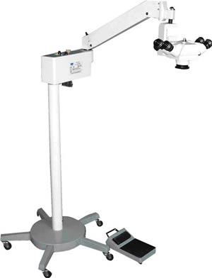
Model XTS―4A
Surgery microscope(Animal microscope)
(operation,surgical
microscope Knowledge,orthopaedics surgical microscope
,Operation,operating)
|
 |
|
|
|
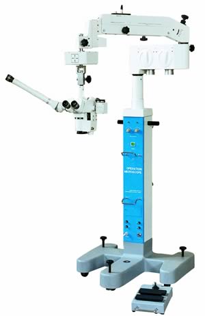
Model LZL―6A Double Binocular
Operation Microscope
(orthopaedics surgical microscope
,Operation,operating) |
 |
|
Getting Paid for New CPT Operating Microscope Code |
Although CPT code 69990
(operation,operating) was
introduced less than a year ago, questions already are
being asked about its proper utilization and how to
obtain reimbursement when the microscope (operation,operating) is used.
The problems arise mainly because many commercial
carriers continue to refuse to pay for microscope
(surgical microscope Knowledge, operation, operating) use,
just as they did before 69990s introduction, when code
61712 (now deleted) was billed for microscope (operation,operating) use in
spinal and brain , while modifier -20 (also deleted,
component) was designated for all other procedures
involving the use of the microscope (orthopaedics
surgical microscope, surgical microscope
Knowledge, operation,operating), according to Susan
Callaway-Stradley, CPC, CCS-P, senior consultant in the
Medical Group at Elliott, Davis and Co., an accounting
and consulting firm in Augusta, GA.
There are a lot of issues regarding this code right now
because not all carriers recognize it, and many coders
are uncertain about usage, she says. Commercial carriers
are beginning to take the stance that if the procedure
is done with a microscope they wont pay for the
use of brain surgery microscopes (operation,operating),Callaway-Stradley
says.
According to Callaway-Stradley, the rationale is as
follows: In the old days, not every hospital had a
microscope, so the old code was used to credit the extra
expense and the expertise required to utilize the
microscope (operation,surgical
microscope Knowledge, operating). But now, virtually all physicians trained to
perform spinal or nerve procedures use a
microscope (operation,operating).
The same is true for ENT procedures, such as
reconstructing the internal ear. You need a scope to
perform those procedures effectively, Callaway-Stradley
says.
In other words, commercial carriers are saying that
since virtually everybody is using the surgery
microscope (operation,operating,orthopaedics
surgical microscope, surgical microscope
Knowledge) and it is an integral part of the procedure
in question, they no longer will reimburse it
separately.
Many 69990 denials have more to do with this trend and
less to do with problems related to the new code itself,
Callaway-Stradley says.
Not For Office Procedures
Commercial carriers aside, there are other issues to
remember when coding for microscope (operation,operating,orthopaedics
surgical microscope, surgical microscope
Knowledge) use. For instance, Medicare, which does
reimburse the use of the microscope (operation, operating), will likely deny a
procedure performed in the otolaryngologists office
using a microscope (operating,operation,surgical
microscope Knowledge) if it was billed with a
69990, because it is supposed to be an add-on code used
during a or procedure.
Note: HCFA will pay about $180, depending on geographic
location, for the 69990, over and above any other
procedure.correct code for cerebral microscope
(operation,surgical microscope
Knowledge,operating) use
during an office procedure is 92504 (binocular
microscopy
( operation,operating) [separate diagnostic procedure]), a special otorhinolaryngologic service code.
Callaway-Stradley says it is difficult, though certainly
not impossible, to get reimbursement for either code,so
before billing for microscope (operation,operating,
orthopaedics
surgical microscope, surgical microscope
Knowledge) use, physicians should
contact their carriers and find out what they require.
This is always good advice, but it is particularly
important with new codes like 69990, and with separate
procedure codes, like 92504, because they might be
bundled into the primary procedure.
Callaway-Stradley calls the 92504 a look-before-you-work
procedure, and says the only time 92504 should be billed
is if it is the only procedure performed on that day. If
the physician uses the microscope (operation,operating,surgical microscope Knowledge,
orthopaedics surgical microscope) to view something during an office visit and
then performs a procedure, the 92504 wont be reimbursed
(unless the carrier is contacted and says otherwise; in
that case, make sure you get it in writing).
In some circumstances, the physician may use the
microscope (operation, operating, surgical
microscope Knowledge) for a purpose unrelated to the primary
procedure. If, for example, during a procedure on the
patients right ear, the physician uses the microscope to
examine the left ear, modifier -59 (separate procedure)
should be attached to the 92504, along with modifier -52
(reduced services).
Multiple Microscope Use
Yet another question about 69990 is whether it can be
billed more than once if the microscope (operation,operating,surgical microscope Knowledge,
orthopaedics surgical microscope) is used for two
or more procedures during an operative session. The
question arises because,with the old codes,physicians
could add modifier -20 to each procedure and get
reimbursed separately for each.Now the question is,do
you get extra reimbursement for each procedure, or just
one flat reimbursement for the entire session?
The answer is entirely carrier specific, according to
Callaway-Stradley, who says she has not seen any HCFA
regulations on the issue. CPT, meanwhile, is unclear on
the subject, though the code book does say 69990 should
be reported in addition to the code for the primary
procedure (operating,operation), which could mean that only the main procedure
should be billed.
The reference to the primary procedure, however, is
vague, Callaway-Stradley says. Some will read it to mean
primary procedure for the session, while others will
interpret it as the main procedure
(operation, operating )with add-on codes.
In the end, Callaway-Stradley says, carriers will likely
interpret it in a way most likely to reduce their costs.
She recommends contacting the individual payer for such
claims and getting the response in writing.
Note: Some procedure codes, such as 31526 (laryngoscopy,
diagnostic, with Operation microscope
(operation,surgical microscope
Knowledge, operating) specifically ban
the use of 69990 in addition to, because the
microscope (operation,operating) is already included. |
| Article Source:http://www.supercoder.com/articles/articles-alerts/otc/getting-paid-for-new-cpt--microscope-code/ |
|
Back To Top |
| |
|
History of the
microscope: from magnifying glass to |
|
Use of the Surgery
Microscope
USE of the operate microscope (operation,operating,surgical microscope Knowledge,
orthopaedics surgical microscope)has now become a sine qua non for good otologic . Effective use of the microscope demands
comfort and in a prolonged this requires not
only practice, but optimal adjustment of the instrument.
If your eyes seem to strain to focus after using the
microscope (operation, operating, surgical
microscope Knowledge) for a few minutes or you are having to
refocus with each change in magnification, it is quite
likely that the optics of your microscope are not
optimally adjusted to you. (It is also possible that you
have a refractive or accommodation error which cannot be
compensated for by the usual adjustments of the
microscope.)(operation,operating,surgical microscope Knowledge,
orthopaedics surgical microscope) |
| Article Source:http://www.ncbi.nlm.nih.gov/pubmed/9574655 |
|
Back To Top |
| |
|
The Efficiency of Operating
Microscope Compared with Unaided Visual Examination,
Conventional and Digital Intraoral Radiography for
Proximal Caries Detection |
Abstract
Objective. The purpose of this study was to evaluate the
efficiency of operation microscope (operation,operating,surgical microscope Knowledge,
orthopaedics surgical microscope)compared with unaided visual examination, conventional
and digital intraoral radiography for proximal caries
detection. Materials and Methods. The study was based on
48 extracted human posterior permanent teeth. The teeth
were examined with unaided visual examination, Operation
microscope (operation, operating, surgical
microscope Knowledge), conventional bitewing and digital intraoral
radiographs. Then, true caries depth was determined by
histological examination. The extent of the carious
lesions was assessed by three examiners independently.
One way variance of analysis (ANOVA) and Scheffe test
were performed for comparison of observers, and the
diagnostic accuracies of all systems were assessed from
the area under the ROC curve (Az). Results.
Statistically significant difference was found between
observers (P<.01). There was a statistically significant
difference between microscope-film
(operation,surgical microscope
Knowledge, operating) radiography,
operating microscope-RVG, unaided visual
examination-film radiography, and unaided visual
examination-RVG according to pairwise comparison
(P<.05). Conclusion. The efficiency of
microscope (surgical microscope
Knowledge,rthopaedics surgical microscope, operation, operating) was found statistically equal with unaided
visual examination and lower than radiographic systems
for proximal caries detection.
1. Introduction
A variety of test methods are discussed for the
diagnosis of proximal tooth surfaces. Adjuncts such as
bitewing radiography and fiber-optic transillumination
provide an improvement to unaided vision. Unaided visual
diagnosis had detected fewer than 50% of caries lesions
on occlusal surfaces and even fewer on proximal surfaces
.
It is not possible to detect only with unaided visual
examination in interproximal caries lesions; radiographs
help for proximal caries diagnosis and detection of
their lesion depth . The combination of visual
inspection and bitewing radiographic images is accepted
as a standard procedure in proximal caries diagnosis .
However, proximal radiolucencies on bitewing radiographs
(operating,operation)
are not always indicative of clinical cavitation. The
deeper the radiolucency penetrates enamel and dentine,
the higher the probability of cavitation .
Due to difficulties in proximal caries detection,
different methodologies were investigated. Magnification
is an accessible, commonly advocated aid to diagnosis .
Recently, the new methods of magnifying visual aids such
as intraoral camera, magnification loops, and Operation
microscope (surgical microscope Knowledge,orthopaedics surgical microscope, operation,operating) are used for caries diagnosis, restorative
treatment decisions, root resection, and retrograde
canal preparation . Previous studies had
investigated the efficiency of operating microscope for occlusal caries diagnosis, but there is insufficient
publication about usage of this device for proximal
caries detection in dental literature.
The purpose of this study was to evaluate the efficiency
of operation microscope (surgical microscope Knowledge,
orthopaedics surgical microscope, operation,operating) compared with unaided visual
examination, conventional and digital intraoral
radiography for proximal caries detection by means of
receiver characteristic (ROC) curve analysis.
2. Materials and Methods
The study was based on 48 extracted human posterior
permanent teeth, 24 molars and 24 premolars stored in a
5% buffered formalin solution. No specimens exhibited
any restoration on the proximal surfaces. Organic and
inorganic debris were removed by an excavator and then
the teeth were cleaned by pumice and water slurry. Three
mouth models were prepared with the teeth to simulate
the clinical condition. The models were fixed in a
phantom head which was adjusted to a dental unit during
the sessions of unaided visual examination and
microscope (surgical microscope
Knowledge,orthopaedics surgical microscope,operation,operating) assessment. The proximal surfaces coronal to
the cementoenamel-junction of the teeth were assessed by
two specialists of oral diagnosis and radiology and one
specialist of restorative dentistry of at least 10 years
of experience independently. To avoid observer fatigue,
an interval of at least one week had separated each
diagnostic session.
The models were examined under a dental unit light, by
using a dental mirror (size 5) and the air water syringe
of the dental unit without any magnification for unaided
visual examination. The clinicians evaluated the extent
of the carious lesions in the proximal surfaces of the
teeth according to a 5-point rating scale (Table 1) .
| Table 1:
Criteria used for evaluations. |
|
|
Scores
|
Visual
examination & operating microscope
|
Radiographic
|
Histological
|
|
|
0
|
No lesion |
Sound |
Sound |
|
1
|
Enamel
opacity with smooth surface |
Radiolucency
in enamel |
Caries in
enamel |
|
2
|
Enamel
opacity with rough surface |
Radiolucency
in dentino-enamel junction |
Caries in
dentino-enamel junction |
|
3
|
Cavitation
restricted to the enamel |
Radiolucency
in the outer half of the dentine |
Caries in
the outer half of the dentine |
|
4
|
Cavitation
extending into dentine |
Radiolucency
in the inner half of the dentine |
Caries in
the inner half of the dentine |
|
|
Then the teeth were
examined using an operation microscope 16x magnification
(Moller-Wedel, Dento 300, Wedel, Germany) according to
the same scale. The observers assessed the teeth
adjusting the height of the stool at a 12
o’clock position. The position of Operation microscope
(operation,operating,surgical
microscope Knowledge,orthopaedics surgical microscope) was not changed to eliminate the position errors during
the examinations. Pictures captured on the computer
monitor were recorded using a video recorder.
After unaided visual and microscope (operation,operating,surgical
microscope Knowledge,
orthopaedics surgical microscope) examinations were
completed, the teeth were mounted in dental stone models
3 in a row (either 2 premolars and 1 molar or 1 premolar
and 2 molars) with proximal surfaces in contact.
Conventional bitewing radiographs of the teeth were
obtained using a specially designed holder to provide
standardized (operating,operation) bitewing projection geometry in the buccolingual direction, tangential to the proximal
surfaces. The object to film distance was approximately
0.5 cm and the source-to-image receptor distance was
32 cm. Size 2 Insight (Eastman Kodak Company, Paris,
France) films with an exposure time of 0.16 seconds and
CCX intraoral unit (Trophy, Instrumentarium, Tuusula,
Finland) with focal spot of size 0.8 mm, at
70 kVp and 8 mA, with 2.5 mm of aluminum-equivalent
(operating,operation)
filtration were used. One centimeter of soft tissue
equivalent material was used to simulate scatter
radiation and beam attenuation from facial tissues. All
film radiographs were developed in automatic film
processor (Velopex, Extra-X, Medivance Instruments Ltd.,
London, UK, and NW107A) with freshly prepared solutions
in the same day.
The film radiographs were assessed using a masked light
box and a 2x magnification X-viewer (Luminosa, CSN
Industrie, Cinisello Balsamo, Italy) by three clinicians
independently in a quiet room with subdued ambient
lighting. Images from the digital system were displayed
on a 17-inch monitor in the same ambient lighting.
Brightness and contrast features of the software were
not changed.The observers (operating,operation) indicated their decision
separately for each interproximal side of the teeth by
masking other side with the use of a black cartoon. They
assessed the extent of the carious lesions according to
a 5-point rating scale (Table 1) .
After all assessments were completed, the teeth were
histologically prepared. The proximal surfaces were
first colored with a solution of propylene glycol with
added basic fucsin (0.5%) for 10 seconds and rinsed in
tap water.Then, the teeth were hemisectioned
perpendicularly to the proximal surfaces from their
santral fossas by a diamond disc under water-cooling. Two sections were obtained, each section was examined
under stereomicroscope (operating,operation) (Olympus SZ 60,Tokyo,Japan) (operating,surgical microscope Knowledge,
orthopaedics surgical microscope,operation)
with a 10x magnification. Two observers not
participating in the study both experienced in
histological examination and being blinded to the
radiographic appearance of the surfaces evaluated the
sections by consensus according to a 5-point confidence
scale (Table 1) .
Histological validation served as a “gold standard” for
all tested methods. One way variance of analysis (ANOVA)
and pairwise comparisons(Scheffe test)were performed
for comparison of observers.The diagnostic accuracies
of the four diagnostic systems were assessed from the
area under the ROC curve (
operating,operation)). Med-Calc (version 7.3) was
used for ROC analysis. The rating scales were
dichotomized as “presence” or “absence” of caries during
the analysis. Score 0 in both radiographic and
histological (operation,operating) scales was detected as absence of caries
and the others were detected as presence of caries.
values were calculated for each observer for each
diagnostic method. The values were analyzed by pairwise
comparison of ROC curves. SPSS-version 13.0 for Windows
was used for all calculations. The level of statistical
significance was α = 0.05.
3. Results
The status of the 96 proximal surfaces of the teeth
were assessed. Histological examination of the teeth
confirmed that 61 (63.54%) of the proximal surfaces were
caries free, whereas 35 (36.46%) of proximal surfaces
determined caries lesions of different depths. The
numbers of proximal surfaces for each score according to
the histological examination are shown in Table 2.
| Table 2:
Histological examination of the teeth. |
|
|
Scores
|
No. of tooth
surfaces |
Percent (%)
|
|
|
Score 0
|
61 |
63.54 |
|
Score 1
|
3 |
3.12 |
|
Score 2
|
12 |
12.5 |
|
Score 3
|
2 |
2.09 |
|
Score 4
|
18 |
18.75 |
|
|
Statistically significant
difference was found between three observers at 99%
confidence interval (P < .01) according to ANOVA.
Scheffe test from pairwise comparisons was performed to
determine which observers were different. No
statistically significant difference was found between
1st and 2nd observers (P < .05) and there was
statistically significant difference between both 1st
and 3rd observers and 2nd and 3rd observers (P < .01)
(Table 3).
| Table 3:
Results of Scheffe test. |
|
|
Observers
|
Groups
|
Mean
difference |
Standard
error |
P
value |
Asymptotic
95% confidence interval |
|
Lower bound
|
Upper bound
|
|
|
1 |
2 |
−0.057 |
0.089 |
.811 |
−0.27 |
0.16 |
|
3 |
0.531(*) |
0.089 |
.000 |
0.31 |
0.75 |
|
2 |
1 |
0.057 |
0.089 |
.811 |
−0.16 |
0.27 |
|
3 |
0.589(*) |
0.089 |
.000 |
0.37 |
0.81 |
|
3 |
1 |
−0.531(*) |
0.089 |
.000 |
−0.75 |
−0.31 |
|
2 |
−0.589(*) |
0.089 |
.000 |
−0.81 |
−0.37 |
|
|
| * The
mean difference is significant at the 0.05
level. |
Two ROC curves are
illustrated. The first ROC curve (Figure 1) is
illustrated by considering assessments of 1st observer
due to no statistically significant difference between
1st and 2nd observers and the second ROC curve (Figure
2) is illustrated for 3rd observer. Areas under the ROC
curve () and standard errors are shown in Table 4 and
analysis of values are shown in Table 5.
| Table 4:
The
Az values and standard
errors for 1st and 3rd observers.. |
|
| |
Test result
variable (s) |
Area
|
Std. error
(a) |
Asymptotic
95% confidence interval |
| |
Lower bound
|
Upper bound
|
|
|
1st Observer
|
Unaided
visual examination |
0.650 |
0.060 |
0.546 |
0.745 |
|
Operating
microscope |
0.650 |
0.060 |
0.546 |
0.744 |
|
Film
radiography |
0.800 |
0.050 |
0.706 |
|
|
RVG
|
0.7933 |
0.051 |
0.698 |
0.869 |
|
3rd Observer
|
Unaided
visual examination |
0.533 |
0.062 |
0.428 |
0.635 |
|
Operation
microscope |
0.533 |
0.062 |
0.429 |
0.636 |
|
Film
radiography |
0.773 |
0.052 |
0.677 |
0.853 |
|
RVG
|
0.760 |
0.054 |
0.662 |
0.841 |
|
|
| Table 5:
Pairwise comparisons of
Az values. |
|
| |
Pairwise
|
Difference
between area |
Std. error
(a) |
P
value |
Asymptotic
95% |
|
confidence
interval |
|
Lower bound
|
Upper bound
|
|
|
1st Observer
|
microscope-unaided visual examination |
0.000 |
0.051 |
.996 |
−0.099 |
0.099 |
|
Operating
microscope-film radiography |
0.150 |
0.072 |
.036 |
0.010 |
0.291 |
|
Operating
microscope-RVG |
0.143 |
0.072 |
.048 |
0.001 |
0.285 |
|
Unaided
visual examination-film radiography |
0.150 |
0.072 |
.038 |
0.009 |
0.291 |
|
Unaided
visual examination-RVG |
0.143 |
0.073 |
.050 |
0.000 |
|
|
Insight-RVG |
0.0077 |
0.054 |
.896 |
−0.099 |
0.113 |
|
|
3rd Observer
|
Operating
microscope-unaided visual examination |
0.001 |
0.036 |
.984 |
−0.070 |
0.071 |
|
Operating
microscope-film radiography |
0.240 |
0.078 |
.002 |
0.087 |
|
|
Operation
microscope-RVG |
0.2266 |
0.078 |
.004 |
0.074 |
0.379 |
|
Unaided
visual examination-film radiography |
0.241 |
0.078 |
.002 |
0.088 |
0.394 |
|
Unaided
visual examination-RVG |
0.227 |
0.078 |
.003 |
0.075 |
0.380 |
|
Film
radiography-RVG |
0.014 |
0.047 |
.772 |
−0.078 |
0.106 |
|
|
For both 1st and 3rd
observers, no statistically significant difference was
found between microscope-unaided
(operation, operating, surgical
microscope Knowledge) visual
examination and film radiography (Insight)-RVG in 95%
confidence interval according to pairwise comparison (P
< .05). There was a statistically significant difference
between microscope-film radiography,
microscope-RVG, unaided visual examination-film
radiography, unaided visual examination- RVG in 95%
confidence interval according to pairwise comparison (P
< .05) for both 1st and 3rd observers.
4. Discussion
The efficiency of microscope (orthopaedics surgical microscope,surgical microscope Knowledge,
operation,operating,) was compared with unaided visual
examination, film and digital intraoral radiography for
proximal caries detection according to ROC analysis in
this study.
Recently, many researchers have advocated the use of ROC
analysis to assess diagnostic methods for the detection
of dental caries . Validity of ROC analysis can be
assessed by increasing the number of tooth surfaces,
increasing the rating scale, and uniform distribution of
caries depths ]. In this study, the sample was
relatively large, 5-point rating scale was used, and the
distribution of caries depths was not uniform. Area
under the ROC curve ( value) gives useful information to
measure accuracy of a diagnostic system . The highest
values belonged to film radiography and RVG for all
observers. The values of unaided visual examination and
Operation microscope (operation,operating,surgical microscope Knowledge,
orthopaedics surgical microscope) were equal and lower than the
radiographic methods.
A diagnostic tool should be reliable and valid.
Interobserver reliability is an important factor for
this aim . On the other hand, training and experience of
observers may affect intra- and interobserver (operation,operating)
agreements. Syriopoulos et al.emphasized that diagnosis
of the radiologists was significantly closer to actual
lesion depth than that of general practitioners. Two of
the observers were the specialists of oral diagnosis and
radiology, the other observer was a specialist of
restorative dentistry of at least 10 years of experience
in this study. No statistically significant difference
was found between the two specialists of oral diagnosis
and radiology for all diagnostic systems (P < .05), but
there was a statistically significant difference between
the specialist of restorative dentistry and the
specialists of oral diagnosis and radiology (P < .05).
The values were found to be 0.800, 0.793, and 0.650 for
film radiography, RVG, and both unaided visual
examination and microscope (operating, operation,surgical microscope
Knowledge,orthopaedics
surgical microscope), respectively,
according to assessments of 1st observer. The values
were found to be 0.773, 0.760, 0.533 for film
radiography, RVG, and both unaided visual examination
and operating microscope (operating,operation,surgical microscope Knowledge,orthopaedics
surgical microscope), respectively, according to
assessments of 3rd observer in this study. The values of
1st observer were higher than 3rd observer for all
diagnostic methods. This condition may be due to the
fact that the specialists of oral diagnosis and
radiology were more experienced than other specialists
about diagnostic and radiographic methods.
Due to difficulty of proximal caries diagnosis with only
visual examination, the combination of visual inspection
and bitewing radiographic images is accepted as a
standard procedure in proximal caries detection [5, 19].
Machiulskiene et al.reported that the clinical
examination alone detected about 60% of the total number
of proximal cavitated dentin lesions, and bitewing
examination detected about 90% of these lesions. But
they emphasized that the clinical examination (operation,operating) is a more
effective method in noncavitated enamel lesions. In this
study, the radiographic methods were better than
clinical examinations for proximal caries diagnosis in
conformity with previous studies .
The positioning of surgical microscope(operating,operation,surgical microscope Knowledge, orthopaedics
surgical microscope) is the most
common difficultness. The operator should be careful and
not change the position as far as possible. It was
reported that the ideal operator zones are in the 7 to
12 o’clock positions for right-handed operators, and 5
to 12 o’clock for left ones. The clinicians should
conform these suggestions to use operating microscope
effectively [22]. The researchers studied at 12 o’clock
position and not changed the position of surgery
microscope (operating, operation,surgical microscope Knowledge, orthopaedics
surgical microscope) during the examinations in this study.
Currently, magnifying visual aids such as magnification
eyeglasses, stereo microscope , and also digital imaging
with magnification are used in proximal caries detection
in some studies and they reported that these methods are
effective. However, Haak et al. reported prism
loupe or microscope ( operating,operation ,surgical microscope
Knowledge, Orthopaedics
surgical microscope) does not improve the
ability to diagnose proximal caries . In this study, the
efficiency of surgery microscope was evaluated by
comparing with unaided visual examination, film and
digital intraoral radiography for proximal caries
detection according to ROC analysis. No statistically
significant difference was found between sur-microscope and unaided visual examination (P < .05), and
there was a statistically significant difference between
Operation microscope (orthopaedics surgical microscope, surgical microscope Knowledge,operating,
operation) and both two radiographic systems
(P < .05).
In conclusion, the efficiency of microscope
was found statistically equal with unaided visual
examination and lower than film and digital intraoral
radiography according to ROC analysis. Because the
surgery microscope (orthopaedics surgical microscope,surgical microscope Knowledge,operating,
operation) is expensive and requires equipment
and operator experience, according to the results of
this in vitro study it can be said that use of this
device would not improve to make an accurate diagnosis
of proximal caries lesions. However, the accuracies of
diagnostic methods with magnifying visual aids should be
investigated and clinical usefulness of these methods in
dental practice should be discussed in vitro and in vivo
with several studies in which the numbers of samples are
larger and rating scales are increased by comparing
conventional methods for proximal caries detection. |
| Article Source:http://www.hindawi.com/journals/ijd/2009/986873.html |
|
Back To Top |
| |
|
|
| An Operating
microscope is an optical microscope
(operation,operating)specifically
designed to be used in a setting, typically to
perform micrurgy.
Design features of a Surgery microscope (orthopaedics
surgical microscope, surgical microscope Knowledge,operating,operation) are:
magnification typically in the range from 4x-40x,
components that are easy to sterilize or disinfect in
order to ensure cross-infection control..
There is often a prism that allows splitting of the
light beam in order that assistants may also visualize
the procedure or to allow photography or video to be
taken of the field.
An example of a procedure which commonly uses a
microscope (operating, surgical
microscope Knowledge,operation) would be endodontic retreatment, where the
magnification provided by the Operation microscope
(operation,operating)
improves visualisation of the anatomy present leading to
better outcomes for the patient. Another example might
be an anastomosis procedure carried out to join blood
vessels in vascular.
Typically an operation microscope (surgical
microscope Knowledge,operating,
orthopaedics
surgical microscope,operation) might cost several
thousand dollars for a basic model, more advanced models
may be much more expensive. Additionally specialized micrology nstruments may be required to make full
use of the improved vision the microscope affords. It
can take time to master use of an microscope
(operation,operating).
Fields of medicine that make significant use of the
microscope include dentistry (especially endodontics),
ENT , opthalmic |
| Article Source:http://en.wikipedia.org/wiki/_microscope |
|
Back To Top |
| |
|
Eye Institute's Dr Mantell
researches Surgery Microscope Damage to the Retina |
During eye ,
neither the patient nor the surgeon thinks much about
the operating microscope(surgical
microscope Knowledge,operating,
orthopaedics
surgical microscope,
operation), except as a mere accessory to
the procedure. Recently, however, I had the opportunity
to do research on the potential dangers of operation
microscopes
(operation,surgical microscope
Knowledge, operating) for an assignment for my Masters. The
results are worth thinking about for all professionals
working in eye care, as well as for patients considering
any eye .
Operation microscopes(surgical
microscope Knowledge,operating,
orthopaedics
surgical microscope, operation) have become an
essential tool in the treatment of ocular disease. It is
well documented, however, that there are risks to the
patient’s eyes from the microscope’s illumination
system. A number of reports have described cases of
retinal damage caused by these microscopes.
The problem is that during , a patient has the
microscope positioned directly above his fully dilated
eye, with his retina typically exposed to the microscope
(operation,operating,surgical
microscope Knowledge, )
light for relatively long periods of OPS.
However, to provide a caveat, damage to the cornea and
crystalline lens from operation microscope (surgical
microscope Knowledge,operating,operation,
orthopaedics surgical microscope) illumination
is always minimised because most of the ultraviolet and
infrared wavelengths in the illumination light are
filtered. The human eye’s exposure limit for these
structures is actually quite high, but it is still
valuable to think about potential damage. The main
concern is potential damage to the retina.
There are many variables that are difficult to quantify,
and each patient is different, so determining the
irradiance in any particular situation is difficult. It
is possible, though, to create useful ‘worst-case’
values that help us determine the risk of retinal
toxicity. These are:
The operation microscope(surgical
microscope Knowledge,operating,operation,orthopaedics surgical microscope) illumination is at maximum
setting.
The pupil is fully dilated.
There is no movement or interruption of the illumination
by eye movement or other factors.
At Eye Institute we take precautions to protect our
patients. We use a modern microscope, which has
appropriate filters to eliminate harmful ultraviolet
radiation.The microscope (surgical microscope Knowledge,operating,operation,
orthopaedics surgical microscope) has advanced optics and this
reduces the need for high illumination to achieve good visualisation. When we always use the lowest
illumination possible and offset the axis so that the
macular is protected. When possible the pupil is covered
to protect the retina. Most surgeries are very short
lasting only 10 to 15 min, this also limits the risk.
Despite the theoretical risks with the above steps the
real risk is extremely low. Reported cases of retinal
toxicity are now extremely rare. |
| Article Source:http://www.eyeinstitute.co.nz/news--microscopes.htm |
|
Back To Top |
| |
|
New System May Supplant
Surgery Operation Microscopes |
Microscopes (surgical microscope Knowledge,operating,operation,orthopaedics surgical microscope) used during are set to face competition in the
future from a new procedure that combines the advantages
of endoscopy and microscopy (operation,operating). Dubbed “Neuro-Comrade,” the
procedure was one of the winners of the Medizintechnik
2006 innovation contest, and its development is to be
funded by the German Federal Ministry of Education and
Research(BMBF) with a project fund of up to €1.5
million for implementation.
In with the Tuttlingen-based endoscope
manufacturer Henke-Sass, Wolf GmbH, a working group
under the leadership of spine specialist Prof. Duffner
has succeeded in taking initial steps towards developing
an intelligent mechatronic restraint system, which takes
over from the previous ergonomically awkward operation
microscope (surgical
microscope Knowledge, operating,operation,orthopaedics surgical microscope) . The mechatronic system operates in
combination with digital imaging and supports the
surgeon’s work. It’s no accident that researchers have
named their system “Neuro-Comrade, since it is designed
to assist the surgeon throughout the entire intervention
by providing precision neuronavigation.
Neuro-Comrade consists of several components. At the
heart is a combination of navigation system and robot
called modiCas. Says Siegen-based project partner Dr.
Jürgen Wahrburg, “We deploy modiCas as a flexibly
controllable restraint system for digital imaging.”
With its optical quality, Neuro-Comrade is expected to
be on a par with conventional microscopes (surgical microscope Knowledge,operating,operation,
orthopaedics surgical microscope) . It is also
expected to enable the creation of multiple pre- and intraoperative images, and allow the operator to control
it simply and ergonomically. |
| Article Source:http://www.devicemed.de/en/industry-news/news/archiv/32_new_system_may_supplant_surgical_microscopes.html |
|
Back To Top |
|