We specialize in
manufacture that Brain surgery microscope .
We sell Brain
surgical microscope etc.�� |
| ��Welcome
to
Surgery
Microscope |
| ��About
Surgical Operating Microscopes |
| ��The
Surgery Microscope |
| ��Split
ear Lobe-Repair under operating microscope |
|
��Operating
microscopes: past, present, and future |
| ��Operating
Microscope Light-Induced Phototoxic Maculopathy After
Transscleral Sutured Posterior Chamber Intraocular Lens
Implantation |
|
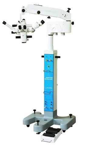
Model LZL-11 Multifunctional Operation Microscope
(Brain surgery
microscope, neurosurgical, Neurosurgery,
Multifunctional,Surgical microscope Knowledge ) |
 ��
|
|
Operating Microscope Light-Induced Phototoxic
Maculopathy After Transscleral Sutured Posterior Chamber
Intraocular Lens Implantation
(neurosurgical,
Neurosurgery,
Multifunctional) |
Purpose: The purpose of
this study is to report the features of surgery
microscope (
Brain surgery
microscope,
neurosurgical,
Neurosurgery,
Multifunctional,Surgical
microscope Knowledge) light-induced retinal phototoxic maculopathy
after transscleral sutured posterior chamber intraocular
lens (TSS PC-IOL) implantation.
Methods: The charts of 118 patients who underwent TSS
PC-IOL implantation at Chonbuk National
University Hospital (Jeonju, Korea) between March 1999
and February 2008 were retrospectively reviewed.
Fourteen patients underwent combined 3-port pars plana
vitrectomy and TSS PC-IOL implantation (vitrectomy
group), and 104 patients underwent TSS PC-IOL
implantation only (nonvitrectomy group). All surgeries
were performed under the same coaxial illuminated
microscope (Brain
surgery microscope,Neurosurgery,Surgical
microscope Knowledge,
neurosurgical,
Multifunctional). All diagnoses were confirmed through careful fundus examination and fluorescein angiography (FA).
Results: Diagnoses of retinal phototoxic maculopathy
were established in 10 (8.47%) of 118 TSS PC-IOL
implantation cases. Phototoxic maculopathy (Brain surgery
microscope,
neurosurgical,
Neurosurgery,
Multifunctional) occurred more
frequently in the vitrectomy group than in the
nonvitrectomy group (6/14 versus 4/104, respectively; P
< 0.001, chi-square = 24.21). Affected patients reported
decreased vision and were found to have coarse
alterations of the retinal pigment epithelium (RPE). In
5 of the phototoxic maculopathy cases (50%), the visual
acuity was 20/200 or worse.
Conclusion: microscope (Surgical
microscope Knowledge,Brain
surgery microscope,
neurosurgical,
Neurosurgery,
Multifunctional) light-induced retinal
phototoxic maculopathy can occur more frequently after
TSS PC-IOL implantation than after casual cataract
, especially when TSS PC-IOL is combined with
vitrectomy. Surgeons should take precautions to
prevent retinal phototoxicity after TSS PC-IOL
implantation and vitrectomy. |
| Article Source: |
|
http://journals.lww.com/Microscope_Light_Induced_Phototoxic.16.aspx |
|
Back To Top |
| �� |
|
Operating microscopes: past, present, and future |
| operating microscope
(Brain
surgery microscope , Surgical microscope
Knowledge,
neurosurgical,
Neurosurgery,
Multifunctional) is a
fixture of modern facilities, and it is a
critically important factor in the success of many of
the most complex and difficult interventions
used in medicine today. The rise of this key
tool reflects advances in understanding the principles
of optics and vision that have occurred over centuries.
The development of reading spectacles in the late 13th
century led to the construction of early compound
microscopes (Brain
surgery microscope , Surgical microscope
Knowledge,
neurosurgical,
Neurosurgery,
Multifunctional) in the 16th and 17th centuries by Lippershey,
Janssen, Galileo, Hooke, and others. Perhaps
surprisingly, Leeuwenhoek's simple microscopes of this
era offered improved performance over his
contemporaries' designs. The intervening years saw
improvements that reduced the spherical and chromatic
aberrations present in compound microscopes (Brain
surgery microscope , Surgical microscope
Knowledge,
neurosurgical,
Neurosurgery,
Multifunctional). By the late
19th century, Carl Zeiss and Ernst Abbe ushered the
compound microscope into the beginnings of the modern
era of commercial design and production. The
introduction of the microscope (Brain surgery
microscope,
neurosurgical,
Neurosurgery,
Multifunctional,Surgical
microscope Knowledge) into the room
by Nyl��n in 1921 initiated a revolution in
practice that gained momentum throughout the 1950s with
multiple refinements, the introduction of the Zeiss OPMI
series, and Kurze's application of the microscope
(Brain
surgery microscope,Surgical microscope
Knowledge,
neurosurgical,Multifunctional,Neurosurgery) to
neurosurgery in 1957.Many of refinements of the
last 50 years have greatly improved the handling and
practical operation of microscope,
considerations which are equally important to
optical performance.Today's sophisticated
microscopes allow for advanced real-time angiographic
and tumor imaging. In this paper the authors discuss
what might be found in the rooms of tomorrow. |
| Article Source: |
| http://www.ncbi.nlm.nih.gov/pubmed/19722819 |
|
Back To Top |
| �� |
|
The
Surgical Operating Microscope |
By: Richard E. Mounce,
DDS
As I travel to lecture and teach, when the surgical
microscope(Brain
surgery microscope, Surgical microscope
Knowledge,
neurosurgical,Multifunctional,Neurosurgery) is discussed, the reasons for its
adoption and/or lack of adoption are uniform.Clinicians who use the surgery microscope (SOM)
state something similar to "It is indispensable. I
cannot practice without it."
Clinicians who do not have the SOM usually state:
1. "I am happy with how I do it. I like my loupes and I
get good results. I don't need it."
2. "Its too expensive. I can't afford it."
3. "I don't want to change my treatment rooms."
4. "I don't want to be slowed down." 5. "It will make me
less profitable."
How can these two positions be reconciled by clinicians
who ideally are honest with themselves (neurosurgical,
Neurosurgery,
Multifunctional) when they are so
mutually exclusive? They cannot.
I make treatment-planning decisions as if the patient
were my wife. If it's good enough for Laura, it's good
enough for all my patients, and vice versa. If the
patient were my wife, I would want her treatment
performed under the SOM. To me anything else is a
compromise, and in the provision(Brain surgery
microscope,
neurosurgical,
Neurosurgery,
Multifunctional) of health care, within
reason, there should be no arbitrarily imposed barriers
to attaining excellence (neurosurgical,
Neurosurgery,
Multifunctional). Simply put, who, being honest
with themselves would want a root canal done with
naked eye or loupes when they could have the same
treatment done under the SOM?
Who do we serve, our patients and the provision of the
best care possible or do we serve our pocket books and
inertia? While at some level that might seem harsh, how
many of us simply cannot afford the $12,000-$15,000 USD
that an entry level SOM would cost? How many of us are
so strapped in our offices that this sum of money
amortized over several years is out of our budgets? I
suspect the answer is very few of us. What then drives
this resistance (Brain surgery
microscope,
neurosurgical,
Neurosurgery,
Multifunctional) ? The simple answer is apathy. We don't
want to change. We are comfortable with how we do it.
The SOM is an abstraction and perceived as an expensive
one. At present, the adoption in North America of SOMs
is approximately 1-5% amongst general practitioners. By
ignoring the value of the SOM, the majority are
literally refusing to see what is clearly there, both
figuratively and literally.
��We are challenged as clinicians to provide our patients
the best possible service while maintaining financial
viability. Part of this challenge is the choice to adopt
the safest and most clinically effective methods.While products
(Brain surgery
microscope,neurosurgical,
Multifunctional,
Neurosurgery) can be debated as to their merits,
visualization and magnification cannot be debated as to
its value. It has inestimable value.
The only people who question the value of the SOM are
most usually those that have never used it. On the other
side of the coin, those that use the SOM state very
clearly that they would never go back to using anything
else. Only the most hardened cynic would argue that SOM
use is superfluous to long-term healing and better
results.
I see the SOM as one component of a larger set of
choices to give my patients the quality of service that
they deserve. For example, in an endodontic (
Brain surgery
microscope,
neurosurgical,
Multifunctional,
Neurosurgery) context,
clinicians have a choice between methods and materials:
they can enlarge canals with Gates Glidden drills and
hand files or they can use the most advanced rotary
nickel titanium (RNT) file on the market today, i. e.
the Twisted File (Sybron- Endo, Orange, CA, USA). They
can use water or only half strength bleach to irrigate
in minimal quantities, or they can use different
irrigants as required and use irrigants that are heated
and ultrasonically activated and use them in sufficient
volume and concentration that the greatest assurance of
cleanliness can be obtained. We can clear the smear
layer (SmearClear, SybronEndo, Orange, CA, USA) or we
can choose to leave a layer of smeared debris, etc. In
essence, there are optimal choices as regards materials
and methods that taken collectively make an immense
impact in the predictability (Brain surgery
microscope,neurosurgical,
Multifunctional,
Neurosurgery) of long term healing. The SOM is one such instrument that in combination with
these other materials and methods define the best we
have to give patients at this time. If we were the
patient, which set of methods and materials would we
want for ourselves?
THE MYTH VS. THE REALITY
Myth #1:
The SOM will slow you down and diminish profitability.Initially, using an SOM will slow things down slightly,
maybe 20% for a few weeks. Initially, there is
awkwardness in using the SOM (Brain surgery
microscope,neurosurgical,
Multifunctional,
Neurosurgery), which ultimately can give
rise to mastery and integration. Gradually, speed is not
affected. There are mutually converging realties that
blend when beginning to use an SOM. One is that the
clinician can see more and hence there is more to do.
For example, if there is a third canal in the mesial
root of a lower molar which may not have been seen with
the naked eye or loupes which is observed under the SOM,
that creates more to do on the part of the clinician, i.
e. another canal system within the tooth to treat.
Alternatively, seeing it allows the clinician to often
know exactly what needs to be done and much of the
guesswork that is present in using tactile feel to guide
root canal treatment is done away with. Locating MB2
canals is far simpler and faster under an SOM relative
to the alternatives (Brain surgery
microscope,neurosurgical,
Multifunctional,
Neurosurgery). More MB2s will be located with the SOM, an event that translates to clinical success, but
also now having to take the time to properly manage the
MB2 will take time.
An added benefit is the full cooperation and
participation of the assistant in becoming central to
the treatment by virtue of being able to ideally see the
treatment on a video monitor as well as to use the
assistant's oculars to obtain the same view as the
clinician.
Myth #2
The SOM is expensive.
Yes, there is an investment, but expensive is dentistry
that has to be redone because it was not performed
correctly the first time. Missing a canal which
ultimately leads to failure of root canal procedure
and subsequent or retreatment is expensive from
two perspectives (Brain surgery
microscope,neurosurgical,
Multifunctional,
Neurosurgery), one is the cost of the procedure that
must be redone and second is the loss of trust which
occurs with the patient looking for another dentist.
Myth #3
The SOM is just for endodontics and doesn't really have
any value in general dentistry.
Nothing could be further from the truth for a general
dentist; the vast majority of procedures could be
performed to a higher standard with the visualization
and magnification provided by the SOM (Brain surgery
microscope,neurosurgical,
Multifunctional,
Neurosurgery), end of story.
From caries detection, to the smoothness of finish
lines, detection of fractures, restoration overhangs,
etc almost every procedure performed can be done more
proficiently with an SOM. As testament to this, it is
virtually unheard of that a clinician purchases an SOM
and then wants to sell it because it doesn't function as
expected. Said differently, the vast majority of the
clinicians (general dentists and endodontists alike) who
buy the SOM are satisfied with their purchases and
utilize the instrument daily.
Myth #4
What I have now is just as good as an SOM, I don't
really need it.If you have this belief, have you looked through the SOM?
Have you ever performed a procedure of any kind under an
SOM?Have you ever been through a microscopic
( Brain
surgery microscope,Surgical microscope
Knowledge,
neurosurgical,
Multifunctional,
Neurosurgery) training
course? Clinicians who are honest with themselves after
these efforts will acknowledge difference between microscopic and the non-microscopic
(Brain surgery
microscope,
neurosurgical,
Neurosurgery,Multifunctional,Surgical
microscope Knowledge) dental world.
Myth #5:
If I get an SOM, I have to get many uneccessary bells
and whistles, like cameras and video equipment and
monitors, etc.
Not true. The entry level G3 Global SOM (Global , St. Louis, MO, USA) has three powers
(magnification levels) and for the clinician that will
never record video or take pictures, this is an
excellent entry level option that has all the
capabilities of much more expensive models, with the
only limit that it possesses three powers relative to a
more advanced model like the G6 which has 6 powers (Fig.
1).
��
HOW TO GET STARTED USING AN SOM
1) Get a loaner SOM on a floor stand and try it out
first before you buy.
2) Take a dedicated SOM training class, even if you
don't have an SOM.
3) Whether you are using a loaner or buy an SOM,
practice your endodontic treatment in extracted teeth
under the SOM extensively and move between the powers
freely using different light intensity to gain
proficiency.
4) Consider joining the Academy of Microscope
(Brain
surgery microscope,Surgical microscope
Knowledge,
neurosurgical,Multifunctional,Neurosurgery) Dentistry, AMED (http://www.microscopedentistry.com) and attending
their annual meeting.
5) Start slowly and be patient, dedicate time to become
comfortable working through the SOM, consider it an
investment in both creating better results as well as
increasing the probabilities of greater profitability
through a higher standard of treatment that will require
fewer "do overs" of all types.
6) Visit other offices that have the SOM and decide
which mounting is ideal for you. Some offices (Brain surgery
microscope,Multifunctional,neurosurgical,Neurosurgery) are ideal
for a ceiling mount, some wall, some swing through, and
some floor stand. Ideally, in the greatest number of
offices the SOM should be mounted, be that on the wall
or the ceiling and not on a floor stand.
7) Talk with other doctors who are using the SOM, get
their feedback as to what works well for them about the
SOM, what they changed in the flow of their office
procedures to accommodate it, etc. For the vast majority
of clinicians, the benefits of the SOM are so persuasive
(Brain surgery
microscope,Multifunctional,neurosurgical,Neurosurgery) that the concept of practicing without it is not an
option.
8) Training resources are available from all the various
manufacturers in the form of literature, course
referrals, in house customer care options, etc.
FAQ:
1) How much does a SOM cost? Entry-level Sums, like the
Global G3 cost approximately $12,000-$15,000 and upwards
depending on the options chosen.
2) If I am not going to record video, give lectures, and
document cases, what do I need as a minimum in an SOM?
Three-step magnification is essential. Having inclinable
binoculars is a nice option but is not absolutely
essential.
3) Which SOM is best to get? I have bought five Global
microscopes (Surgical
microscope Knowledge,Brain
surgery microscope,neurosurgical,Multifunctional,Neurosurgery) in my practices over the years and have been
very satisfied. Global is an excellent option for
quality at the price point. Other brands may be more or
less expensive, but it must remembered that an SOM is a
one-time expense and a lifetime purchase, they do not
wear out, repair issues are rare. Global SOMs are
modular and expandable as desired, in other words if you
want to add video or cameras later, they can be added
easily.
4) How much of the procedure do I do under the SOM? How
do know when I should be looking under the SOM and when
I don't have to?
In my treatment rooms, the SOM is my overhead light
source, I do not have a special dedicated (Brain surgery
microscope,neurosurgical,
Multifunctional,
Neurosurgery) light source
and the SOM. I use the SOM for everything from access to
occlusal adjustment. At no time in the process do I work
without the SOM. It is indispensable for me (Figs. 2-5).
A discussion of the value of the operation microscope
(Surgical
microscope Knowledge,
Brain surgery
microscope,neurosurgical,Multifunctional,Neurosurgery) has been presented with a view to making
clinicians aware of its capabilities and improvement in
the quality of care possible.
Dr. Mounce is on the advisory council of SybronEndo and
is paid for some aspects of this position, lecturing for
example, otherwise he has no commercial interest of any
kind in the materials or methods described in this
paper. Dr. Mounce offers intensive customized endodontic
(Brain
surgery microscope,Surgical microscope
Knowledge,
neurosurgical,
Multifunctional,
Neurosurgery)
single day training programs in his office for groups of
1-2 doctors.For information,contact Dennis at
360-891-9111 or write RichardMounce@ Mounce Endo.com. Dr.
Mounce lectures globally and is widely published. He is
in private practice in Endodontics in Vancouver, WA, USA |
| Article Source: |
| http://www.oralhealthjournal.com/issues/story.aspx?aid=1000223095&type=Print%20Archives |
|
Back To Top |
| �� |
|
Split ear Lobe-Repair under operating microscope |
Since ancient times, men
and women in all civilization have been adorning their
ears with earrings (neurosurgical,
Neurosurgery,
Multifunctional) for ornamental ad cultural reasons��
Over the years, the tradition of earrings in India has
strengthened and today it is customary for women in most
communities to have their ears pierced from an early
age.
Torn earlobes
(Brain
surgery microscope, Surgical microscope
Knowledge,
neurosurgical,
Multifunctional,
Neurosurgery) are one of the most common problems
effecting people with pierced ears. Earlobes may have
enlarged holes or be split as a result of heavy earrings
or pulled earrings. The increasing practice of ear
piercing has resulted in more and more requests for ear
lobe repairs.
Methods of reconstruction
Split ear lobe repair under an operating microscope
(Surgical
microscope Knowledge,Brain
surgery microscope,neurosurgical,Multifunctional,Neurosurgery)
Depending on the deformity, reconstruction can take
different forms. In all methods, the skin lining the
slot is removed creating a raw edge to rebuild. Lost
tissue complicates matters and reconstruction centers
around reestablishing normal proportions in a somewhat
smaller ear.
Layered closure using fine suture materials under local
anesthetic is performed. Either straight line closure or
z pasty can be done to reduce the scar line.Scar formation and keloid tendency is to be born in mind
before embarking on the procedure (Brain
surgery microscope,Multifunctional,
Neurosurgery,Surgical microscope
Knowledge,neurosurgical) . An informed consent
in this regard is taken before.
Most patients should be explained about the possible
scar line, and delay before wearing another ear ring or
re piercing (minimum six weeks).They should be told to
take extra care not to get the wound infected, avoid UV
light while going out.
A cosmetic make up with UV sunscreen will camouflage the
scar for the six weeks period. Or start wearing a clip
on ear stud after 3-4 weeks before re piercing.
Postoperative Care
A thin layer of antibiotic (Brain
surgery microscope,Multifunctional,
Neurosurgery,Surgical microscope
Knowledge,neurosurgical) ointment (Neosporin®) is the
only dressing needed in most cases. You may wash your
hair after putting the ointment.
The sutures are removed in 7 days.
Re piercing (neurosurgical,
Neurosurgery,
Multifunctional)
Wait minimum of six weeks, No compromise on this, even
if the patient becomes ��impatient��
Clip on earrings may be worn 3-4 weeks after
How to avoid scar and keloid-Scar care(Brain
surgery microscope,Multifunctional,
Neurosurgery,Surgical microscope
Knowledge,neurosurgical)
After sutures are removed, one can start rubbing
Contratubex® gel twice a day for a month or two. It will
prevent thickened scar formation to a great extent.
The active ingredients in Contratubex® are cepae
extract, heparin and allantoin.
• Cepae extract is obtained from onions. It generates an
anti-inflammatory, bactericidal effect (neurosurgical,
Neurosurgery,
Multifunctional). And it reduces
swelling while preventing excessive growth of the
connective tissue.
• Heparin loosens the tissue structure. It has an
anti-inflammatory effect and helps to bind water to the
scar tissue.
• Allantoin encourages wound healing and has a soothing
effect. In older scars its most important effect is to
replenish and regulate the extreme lack of water in the
scar tissue �C and to promote blood flow
Using Operation microscope(Surgical
microscope Knowledge,Brain
surgery microscope,
neurosurgical,Multifunctional,Neurosurgery) for Split ear lobe repair
Occasionally I use surgery microscope to repair the
split ear lobe, in ��costomers�� who are very demanding
and want perfect result, often coming from the upper
class.
But I must admit, the results are much better when I
used the microscope (Brain
surgery microscope,Multifunctional,
Neurosurgery,Surgical microscope
Knowledge,neurosurgical), although takes more time ,
and not always easy when I have got a tight schedule,
with other more important procedures waiting. |
| Article Source: |
| http://www.drpaulose.com/general/split-ear-lobe-repair-under-microscope |
|
Back To Top |
| �� |
|
Welcome to
Surgery Microscopes
(neurosurgical,
Neurosurgery,
Multifunctional) |
| The invention concerns a
microscope having a power and data transfer system
between a microscope body and an external control device
or peripheral device. The invention relates generally to
surgical microscopes (Surgical
microscope Knowledge ,
Brain surgery
microscope,neurosurgical,Multifunctional,Neurosurgery), and particularly to an
improved configurations for linking a microscope body to
an external power supply, control device, and light
source. A surgical microscope for the purposes of the
invention is understood to be a microscope(Surgical
microscope Knowledge,Brain
surgery microscope,neurosurgical
,Multifunctional,Neurosurgery) that is
movable with respect to an object and thus possesses
certain flexibility in terms of any connections to
external devices. Such microscopes are very often used
in operations. Such microscopes are often also
used for industrial or commercial applications.Such
microscopes (Multifunctional,
Neurosurgery,
Brain
surgery microscope,
neurosurgical) often have an integrated illumination system
in which the light source is built into the microscope.
With such external accessories, the light is directed
through a light guide from the external light source to
the microscope (Surgical
microscope Knowledge,Brain
surgery microscope,neurosurgical, Multifunctional, Neurosurgery) body, and through the latter onto the field. Such microscopes are often located on
the extension arms of stands, while the external devices
and control systems are housed in the column region of
the stand. The connection between the external devices
and the microscope body or the terminals located thereon
is accomplished via flexible lines such as light guides,
electrical cables, electronic data lines, etc. In some
cases they interfere with visibility, are heavy, result
in jamming and limitations of movement, and moreover
look untidy. In the field of surgical microscopy
(neurosurgical,
Surgical microscope Knowledge,Brain
surgery microscope,Multifunctional,Neurosurgery), they
result in increased surface areas which thus make the
overall structure more susceptible to soiling. The
assignee of the present application has already taken
initial steps intended to remedy this unfavorable
situation. This hose was relatively bulky and
inflexible, however, and did not make optimum use of
space since it had to be made sufficiently large for
subsequent installation of an undetermined number of
cables, even if not all the cables were pulled through.
It is thus the object of the invention to implement the
connection between the external devices and the
microscope (neurosurgical,
Surgical microscope Knowledge,
Brain surgery microscope,Multifunctional,Neurosurgery) body in as lightweight, easily movable, and
retrofit table a fashion as possible, and with as few
cables as possible. The present invention, as broadly
defined, achieves this principal object on the basis of
a physical size reduction and simultaneous weight
reduction. A preferred configuration of a cable
according to the present invention, which optionally can
also be used independently of the invention, is
coaxially multi-layered, one of the layers, but
preferably the core of the cable, being configured as a
mirror optical system or fiber optical system or as a
liquid light guide, while at least two layers are
configured as an at least two pole power cable.
Identical part bear identical(neurosurgical,surgical
microscope Knowledge,Brain
surgery microscope,
Multifunctional,Neurosurgery) reference characters;
different parts having functions that are identical in
principle bear identical reference characters with
differing indices. A power connection 4c in the form of
a light guide transmits power in the form of light flux.
The microscope (neurosurgical,
Surgical microscope Knowledge,Brain
surgery microscope, Multifunctional,Neurosurgery) thus comprises a terminal 3 for power
connection 4c and a terminal 6 for the data connection.
An extremely wide variety of combinations lies within
the context of the invention. This includes the case in
which electrical signals are transferred over the light
guide by light modulation. The invention encompasses, on
the one hand, corresponding modulation of the electrical
(neurosurgical,
Surgical microscope Knowledge,
Brain surgery microscope,Multifunctional,Neurosurgery)
or light fluxes that are flowing in the manner of power,
and/or the fact that electrical or optical signals are
sent, parallel to these flow power fluxes, over the same
line in each case. |
| Article Source: |
| http://www.surgerymicroscopes.com/ |
|
Back To Top |
| �� |
|
About Surgery Microscopes
(neurosurgical,
Neurosurgery,
Multifunctional) |
We have created this
website to provide useful and significant information
between our customers about the various types of
microscopes (neurosurgical,
Surgical microscope Knowledge,Brain
surgery microscope,Multifunctional,Neurosurgery)that we sell, specifically the
operating microscopes. We create this informative
website to help our customers and our frequent readers
get a better understanding about the world of
microscopy, various types of microscopes but more
specifically surgical operation microscopes
(neurosurgical,
Surgical microscope Knowledge,Brain
surgery microscope,Multifunctional,Neurosurgery)and other topics and
issues related to operating microscopes.
We strive hard and do our best to provide you a good
reference material on surgical microscopes. It
is our intent for this science reference website to be
devoted to the subject matter and provide useful
articles to the students, teachers, professionals and to
everyone that are interested in microscopy
(Surgical
microscope Knowledge,neurosurgical,
Brain surgery microscope,Multifunctional,Neurosurgery) related
subjects. We research diverse sources of related and
appropriate information to create comprehensive articles
about surgical operation microscopes for our website.
We make sure that every article we upload to our website
about the surgical microscopes are relevant to
the subject matter. We have a team of well-qualified
article writers, editors and researchers that offer full
interests to their work in creating useful and
comprehensive articles about surgical operating
microscopes (
Multifunctional,Neurosurgery
,Surgical microscope Knowledge,
neurosurgical,
Brain surgery microscope) . They are the ones who also make sure that
the text content on our website are accurate to avoid
misinformation between our readers. Feel free to check
out our website to get the latest deals about the
operating microscopes. You can contact us and
talk to one of our technical support on microscopes for
any of your microscope (neurosurgical,
Surgical microscope Knowledge,
Brain surgery microscope,Multifunctional,Neurosurgery) needs.
The success of
endodontics relies on the localization of the entire
root canal system and its subsequent cleaning, shaping,
and three-dimensional obturation. Several magnification
systems have been advocated over the years.
The most convenient and popular have been loupes with
varying degrees of magnification. However, the
introduction of the surgical operating microscope
(neurosurgical,
Surgical microscope Knowledge,
Brain surgery microscope,Multifunctional,Neurosurgery) (SOM)
to endodontics has dramatically changed the practice of
the specialty.
In endodontics, the introduction of the SOM is
relatively new concept that is revolutionizing the way
procedures are performed.
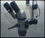
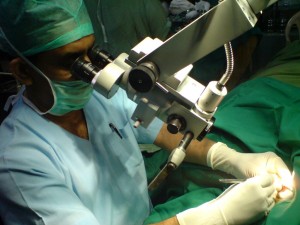
The SOM with video camera attached. The degree of
magnification depends upon the combination of lenses
that are utilized. Most microscopes
(Surgical
microscope Knowledge,neurosurgical,Brain
surgery microscope ,
Multifunctional,Neurosurgery) come with three to
fives steps of magnification ranging from 3x to 27x. The
light source is usually 100 to 150 watt halogen bulb
that is connected to the microscope via a high
efficiency fiber optic cable.
Many other options may be added to the microscope
(Surgical
microscope Knowledge,
neurosurgical,
Brain surgery microscope,Multifunctional,Neurosurgery) such
as an assistant's viewing eyepiece, video, and 35mm
cameras. These options provide a very powerful tool that
can be used for teaching, patient education, and medical
legal purposes. |
| Article Source: |
| http://operatingmicroscopes.com/about.html |
|
Back To Top |
|