We specialize in
manufacture that operating microscope
for Ophthalmic .
We sell
operatingmicroscope for Ophthalmic etc.
|
| ■Operating
Microscopes in Ophthalmic Surgery |
| ■Surgical
Microscope |
| ■Surgical
Microscopy
and Dentistry |
| ■Public
Health Advisory: Retinal Photic Injuries From Microscopes
During Cataract Surgery |
| ■Inventions:
Frameless Stereotactic Operating Microscope |
| ■General
Facts, Ways, and Tips on How to Use a
Microscope |
|
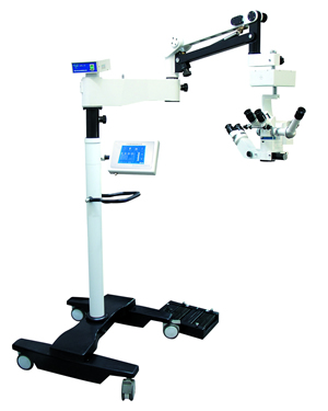
Model LZL-16
Operation microscope for ophthalmology
(Ophthalmologic,
Ophthalmic) |
 |
|
Public Health Advisory: Retinal Photic Injuries From
Operating Microscopes During Cataract Surgery |
This message from the
Food and Drug Administration (FDA) is to remind you
about the potential retinal hazards from surgery
microscopes (Ophthalmologic,
Ophthalmic), to review steps that can be taken to
minimize the risks of retinal photic injury from
operation microscopes (Ophthalmologic,
surgical microscope,Operating
Microscope,Ophthalmic,Operating Microscope Knowledge ), and to make sure physicians are
aware of medical device reporting.
Background
Retinal photic injury from an microscope (ophthalmic ,surgical microscope ,Operating Microscope Knowledge,ophthalmologic) was
first reported in 1983. (1) Since that time, incidents
of retinal photic injuries from surgery microscopes (Ophthalmologic,Ophthalmic),
continue to be reported sporadically. (2-16) While the
majority of injuries produce minimal symptoms, scotoma
and permanent central vision loss have occurred in some
patients. (11) As is true with all photochemical damage,
clinical abnormalities are not evident immediately.
Retinal edema or mild pigmentary changes are typically
discernable within one to two days after exposure and
varying degrees of pigmentary modeling become more
apparent after one to three weeks. (18)
Incidence of Injuries
The incidence of serious injury is not known, as is
illustrated by the following studies, but significant
permanent vision loss appears to be infrequent. A recent
prospective study at a training institution found
retinal photic injury from a surgery microscope (Ophthalmologic,
surgical microscope,Ophthalmic,Operating Microscope Knowledge ) in
28% of patients. (11) In this study a microscope was
used with relatively intense light and exposure times of
20 to 120 minutes. This study also demonstrated a
dose-response relationship; the risk of retinal damage
increased with increasing retinal exposure to the light
from surgical microscopes (ophthalmologic,ophthalmic).
Another prospective study, performed at a different
institution with an operation microscope (Ophthalmologic,
surgical microscope,Operating
Microscope,Ophthalmic,Operating Microscope Knowledge )that provided
about 28% lower corneal irradiance levels and shorter
exposure times (21 to 76 minutes), reported no retinal photic injuries. (16) Preoperatively, patients had
detailed ocular examinations and , when possible, fundus
photography and oral fluorography. Fluorescein
angiography was performed after cataract to
identify the most subtle retinal photic injuries that
may have occurred. In two additional retrospective
studies, the incidence of retinal photic injuries was 7%
and 3%. (8, 17)
Risk Factors
Despite all efforts taken to minimize the risks of
retinal damage, retinal photic injuries from the light
source used in surgery microscopes (ophthalmologic,ophthalmic) during cataract and other intraocular procedures may occur.
Several factors appear to be important determinants of photic retinal injury. These include: angle of light
incidence, light intensity, exposure time, and intensity
of the blue light component.
Actions to Reduce the Risk of Retinal Photic Injury
The following actions may reduce the risk of retinal
photic injury from surgery microscopes (ophthalmologic,ophthalmic) during cataract
ops:
・Use
only that light intensity needed to clearly visualize
and perform the procedures.
・Do
not assume that the intensity of the light from all
surgery microscopes (Ophthalmic,
Ophthalmologic, surgical microscope,Operating Microscope Knowledge) is the same. Some are brighter
than others. When using a new microscope(Ophthalmic,Ophthalmologic), visually
evaluate and set light levels to the lowest levels
successfully used in the past.
・Replace
lamps only with manufacturer-approved products.
・Because
blue light has been shown to be more toxic than
longer-wavelength light, the addition of a filter to
exclude light below about 515 nm has been recommended,
to eliminate blue light, especially in cases requiring
prolonged light exposure. (18) However, a 515 nm short
wavelength cut-off filter will result in a yellow light.
Cut-off filters at wavelengths shorter than 515 nm to
about the range of 420 - 435 nm will affect the color
rendition of the light less and may still provide useful
reduction in the risk of injury.
・Use
oblique lighting if it is available, or otherwise shield
the pupil when the red reflex is not required or the field permits. Oblique lighting may be used
during phases of an that do not require
coaxial light.
・Minimize
direct exposure to the fovea.
・Educate
residents about the above actions in order to help
reduce the risks of retinal photic injury during
training programs and in the future.
Standards Efforts
The American National Standards Institute (ANSI) is
developing a proposed product performance standard that
will be applicable to the manufacturers of surgery
microscopes (ophthalmologic,ophthalmic) used in ophthalmic surgery. This proposed
standard will incorporate engineering, labeling, and
user information requirements that are intended to make
the device safer to use and the user aware of ways to
minimize the risks associated with the use of the
surgical microscope (Ophthalmic,Ophthalmologic) during ocular OPS.
Reporting Requirements for Retinal Photic Injury
Incidents
Although retinal photic injury from surgery
microscopes(Ophthalmic,
Ophthalmologic, surgical microscope,Operating
Microscope,Operating Microscope Knowledge) during cataract is not a new
phenomenon, some physicians may not be aware of the
reporting requirements of the Safe Medical Devices Act
of 1990 (SMDA). Prompt and accurate reporting by
practitioners will help make it possible to obtain a
better estimate of the incidence of retinal photic
injury from surgical microscopes (ophthalmologic,ophthalmic) during cataract and other intraocular procedures.
The SMDA requires hospitals and other user facilities to
report deaths, serious illnesses and injuries associated
with the use of medical devices. The procedures
(Ophthalmologic, Ophthalmic)established by your facility for such mandatory
reporting should be followed. Practitioners who become
aware of any medical device related adverse event or
product problem/malfunction should report to their
Medical Device User Facility Reporting person.
Even if an incident is not required to be reported under
the SMDA, it would be helpful to report directly to
MedWatch, the FDA's voluntary reporting program. |
| Article Source: |
| http://www.fda.gov/MedicalDevices/Safety/AlertsandNotices |
|
Back To Top |
| |
|
Inventions: Frameless Stereotactic Operating
Microscope |
Inventors: Professor
John Strohbehn and Dr. David Roberts DMS’75
Ever since early humans drilled holes into patients’
heads in paleolithic neuro, doctors have longed
for a way to navigate the brain and pinpoint lesions. In
the 1970s computerized tomography (CT) produced (Ophthalmologic,Ophthalmic)amazing
two-dimensional images of the brain, but the only way to
use the scans as navigational guides during was
via a cumbersome metal frame that ringed the patient’s
head, got in the surgeon’s way, and (ouch!) had to be
screwed directly into the skull.
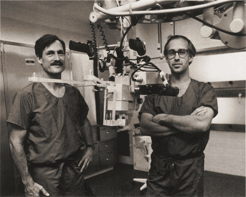
In the early 1980s Dartmouth-Hitchcock Medical Center
neurosurgeon David Roberts DMS’75 asked Thayer Professor
John Strohbehn to create a better solution: an
instrument that could map CT data onto the visual field
of a microscope(Ophthalmic,Ophthalmologic)to produce a precise three-dimensional
(a.k.a. stereotactic) view of the brain. Working
together in Strohbehn’s lab at 7 a.m. ― before Roberts’
clinical hours and Strohbehn’s classes ― they created an
operation microscope (Operating
Microscope Knowledge, Ophthalmic, Ophthalmologic,
surgical microscope) that was stereotactic, frameless,
and precise. They tested their prototype in the
room in 1983 and patented the invention three
years later.
The frameless stereotactic operating microscope (surgical
microscope,Ophthalmologic, Operating Microscope
Knowledge, Ophthalmic, Operating Microscope) was a
hit. Not only was it more comfortable for the patient,
it was the beginning of image-guided .
Today every room in the world is
equipped with an updated version of Strohbehn and
Roberts’ invention. You don’t have to be a brain surgeon
to know that brain would now be unthinkable
without it. |
| Article Source: |
| http://www.dartmouthengineer.com/2007/05/inventions-spring-2007/ |
|
Back To Top |
| |
|
General Facts, Ways, and Tips on How to Use a
Surgical Operating Microscope |
Surgery is a branch of
medicine that deals with the treatment of deformities,
injuries and diseases through a series of procedures and
techniques. It has become the main solution for people
with physical problems. It allows man to remove and
replace damaged organs and tissues, fix broken bones,
heal vital parts of the body, and reconnect limbs caused
by accidents. The influences caused by surgical
microscopes (Ophthalmic,
Ophthalmologic, surgical microscope, Operating
Microscope Knowledge) in the fields of
OPS and other medical research have provided
surgeons to perform seemingly impossible tasks. These
microscopes (Ophthalmic,Ophthalmologic) allow the surgeons to observe and examine
the part of a body to be operated. Some of these body
parts are too small to be seen with the naked eye such
as blood vessels, nerve fibers and organ tissues. An needs careful handling and a scrutinized
examination which is why it is impossible to perform
without this microscope(Ophthalmic,Ophthalmologic). Patients who have undergone today are contented with the results. During
the past, undergoing makes a
patient cower in fear because the risks he or she might
have but with today’s newest developments, a failed
operation is less likely to happen.
Definition of a surgical microscope
This microscope is a type of binocular microscope (Ophthalmologic,Ophthalmic), which
enables the surgeon to get a detailed view on the
structures of an area going through a procedure.
Also known as an microscope(Operating
Microscope,Ophthalmic,
Ophthalmologic, surgical microscope, Operating
Microscope Knowledge), it is
equipped with a motorized zoom lens system which
provides the person to perform his task at a good
working distance and a set of exchangeable oculars which
offers different magnifications.
Parts of a Surgical Operating Microscope
operation microscopes (surgical microscope ,
Operating microscope Knowledge, ophthalmic,Ophthalmologic)
have varying capabilities, depending on their type of
use. They, however, share some basic common features.
The first common feature is a heavy movable stand to
ensure its stability which has an attached arm that
allows for manipulation of the optical portion to be
directed to the area. The optical head has one or
two pairs of binocular eyepieces and a high intensity
light source. Halogen-tungsten lamp and fiber-optic
coaxial illumination act as light source of the microscope
(Ophthalmologic,
Ophthalmic). Foot pedal power control over zoom, focus,
light, and position are also present in more
sophisticated microscopes. Generally,a good surgical
operation microscope (ophthalmologic,ophthalmic, Operating
Microscope, surgical microscope, Operating
Microscope Knowledge) should have excellent optics, motorized
focusing, floor stand mounting on wheels for easy
portability, and coaxial illumination.
How to use a surgical operation microscope (ophthalmologic,ophthalmic)
In using this microscope (Ophthalmologic,Ophthalmic), there are several ways and
tips to know to get good quality images on the part
where the is taking place. Here are some
things of how to use this tool.
•
All valuable things must be
handled with care. A surgical operation microscope
(surgical microscope,
Ophthalmologic,Operating Microscope,
Ophthalmic,Operating Microscope Knowledge) is a
vital tool included in OPS, therefore, the user must
ensure that it must be in its best state during the
whole procedure. Lack of attendance may lead to a
deficient performance. In addition, keeping a regular
check on the components of this microscope(Ophthalmologic,
Ophthalmic) should also
be done. Constant use of this tool could also add to a
decline on its performance so replacements must always
be ready.
•In
order to get a good view of the operated area,adjusting this microscope
(ophthalmologic, Ophthalmic) to high contrast is necessary.
Having a high contrast could make the user carry out his
work precisely. Also, avoiding light reflections
improves the contrast so it must be noted that this
microscope(ophthalmologic,ophthalmic) must always be in its completely sealed
housing.
•To
easily move this microscope (ophthalmologic,ophthalmic) from one position to
another, adjusting its prisms helps without affecting
the capability to obtain images of impressive quality.
•
When performing a OPS, the surgeon’s hands must
focus on the of the patient rather than
concentrating on the controls of this microscope (Ophthalmologic,
Ophthalmic).
Therefore, making use of its features such as its
motorized control with the use of the surgeon’s foot
greatly helps.
•Modern
gadgets attached to it must also be used to help the
surgeon in his A motion-picture camera makes
it easy to record the functions and activities of the
for documentation purposes and for references.
•
The halogen lamp attached to this tool provides the
surgeon to obtain the particular image without
difficulty. As long as it is well-focused, there is no
need to provide a secondary light source anymore.
•Keeping
it dry and clean must also be observed particularly on
the most essential components such as its objective
lenses and oculars.
•
After using it, it must be kept on a secured container
that could protect it from getting wet, heat, and other
physical damages it might undergo.
Being a surgeon is indeed a valued profession. The
accomplishments he or she achieves result from the
dedication of work, the techniques and the tools he
used, one of which is the surgical microscope (surgical
microscope, Ophthalmologic,Operating Microscope,
Ophthalmic,Operating Microscope Knowledge).
A lot of people whose lives were endangered and have
undergone are saved. Many are also thankful that
they live a more comfortable life after going through an |
| Article Source: |
| http://www.microscope.com/general-facts-ways-and-tips-on-how-to-use-a---microscope.html |
|
Back To Top |
|