|
We specialize in
manufacture that operating microscope for
Ophthalmic.
We sell operating
microscope for Ophthalmic (ophthalmology) etc.
|
| ■Operating
Microscopes in Ophthalmic Surgery |
| ■
Operating Microscope |
| ■Surgical
Microscopy
and Dentistry |
| ■Public
Health Advisory: Retinal Photic Injuries From Operation Microscopes
During Cataract |
| ■Inventions:
Frameless Stereotactic Operating Microscope |
| ■General
Facts, Ways, and Tips on How to Use a
Operation Microscope |
| |
|
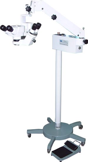
Model XTS―4C Microscope For Ophthalmic Operation
(Operation
Microscope,Ophthalmic
surgical microscope,
ophthalmological,
Ophthalmologic,ophthalmology
)
 |
|
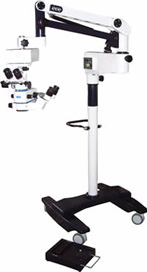
Model LZJ―6D
Operation Microscope for Ophthalmologic Operations
(Ophthalmologic,Operation
Microscope,Ophthalmic
surgical
microscope,
ophthalmological,
ophthalmology )
 |
|
Operating Microscopes in Ophthalmic Surgery |
Problem
Exposure to light from an operation microscope(Operation
Microscope,Ophthalmic
surgical microscope,Ophthalmological,Ophthalmologic,Ophthalmology)during extracapsular cataract extraction (ECCE) and intraocular
lens (IOL) implantation can result in photic retinopathy
(light-induced retinal damage), particularly in patients
with existing retinal impairment. ECRI investigated
several incidents in which eye lesions occurred in
patients who underwent ECCE with IOL implantation. The
shape and orientation of these lesions were consistent
with those that would be expected from retinal exposure
to excessive light intensity from a fiberoptic, focused
delivery system. Contributing factors were prolonged
exposure and high illumination levels. In an unrelated
series of incidents, 11 separate lawsuits were filed in
1985 against a hospital in connection with eye injuries
allegedly resulting from use of an operation microscope ( Operation
Microscope,
Ophthalmological,
Ophthalmic
surgical microscope,Ophthalmology)
with a missing heat filter for ophthalmic (Ophthalmology) surgery.(1)
Discussion
Clear visualization of the field is essential
for successful . To enhance the surgeon's view of
microscopic (Ophthalmic
surgical microscope,Operation
Microscope,
Ophthalmological,
Ophthalmology) structures(e.g,nerves,blood and
lymphatic vessels,lesions),magnification is necessary. An surgery microscope
(Ophthalmological,Ophthalmology) is needed for procedures in
which the surgeon requires adjustable focusing
capability and greater stability than offered by a
loupe.
When an operating microscope (Ophthalmic
surgical microscope,Operation
Microscope,
Ophthalmological,Ophthalmologic,Ophthalmology) is used, light is
transmitted from a lamp housing into surgery microscope's(ophthalmological,ophthalmology)body through prisms or fiberoptic cables and is
transmitted through the objective lens to the
field, concentric to the field of view. The light
reflected from the field passes through the
objective lens, the magnification changer, and the
eyepieces, where the surgeon sees the image of the field. When the patient's eye is exposed to
intense light (especially short-wave or near-ultraviolet
wavelengths)for extended periods,problems such as cystoid macular edema and postprocedural vision blurring
may result. Photic retinopathy should be considered when
vision blurring persists (Ophthalmology,Ophthalmological) following ECCE. Such blurring
usually clears up in less than two months; however, in
1-2% of all ECCE patients it does not.
The complex interaction of the many factors that affect
retinal light exposure has prevented standards for
exposure limits from being established. Although
illumination must provide a clear field of
view for the surgeon, the intensity of illumination on
the field depends on the surgeon's own visual
acuity and the efficiency of the operation microscope (Ophthalmic
surgical microscope,operation
Microscope,ophthalmological,ophthalmology) to
transmit reflected light back from field.The
duration of illumination is also an important factor in
retinal damage.Thus,different surgeons and procedures
will subject the eye to varying levels and durations of
exposure.Additional variable factors such as
anesthesia,light heat filters,and corneal
protective devices(e.g,eclipse filters, semiopaque
contact lenses) must also be taken into consideration.
The only recommendations that can be made are
cautionary. Even when precautions are taken and users
are properly trained, retinal damage is a recognized
risk. Phototoxic retinal exposure can be reduced by
limiting overall level and time of illumination,
irrigating the eye frequently,and covering the central
cornea with a semiopaque contact lens or alternative
method before lens extraction and during wound closure.
Side-beam illuminators and IR/UV and yellow light
filters are recommended to decrease light exposure
levels. Most manufacturers offer these filters for their
microscope (ophthalmologic,Ophthalmic
surgical microscope,
operation Microscope,ophthalmological,ophthalmology) systems.
Recommendations
Alert all ophthalmic surgeons to this report and keep
them informed of the most recent changes in user
instructions issued by surgery microscope (Ophthalmologic,Ophthalmic
surgical microscope,operation Microscope,ophthalmological,ophthalmology) manufacturers.
Surgeons should be up to date on any microscope system
upgrades, additional safety features, changes in patient
selection protocol as outlined by the manufacturer, and
procedural changes.
Use the lowest light intensity setting at which
acceptable field visualization can be
achieved. Consistent with clinical needs,surgeons
should not decrease the aperture for added depth of
field. The benefits of the added depth may be outweighed
by the risks of the additional light needed for field
visualization.
Always use protective corneal covers except when full
visualization of the anterior chamber is necessary, and
keep the eye properly irrigated at all times.We recommended the use of side-beam illuminators and
safety filters to reduce the risk of patient injury.
Surgeons should be knowledgeable in their proper use.
Add a safety check to the preoperative checklist to be
sure that all such devices are installed and functional.
Note
Missing heat filter triggers suit. Malpractice Rep
1985;(Jun):10.
UMDNS Term
Microscopes (Ophthalmologic,Ophthalmic
surgical microscope,
Operation Microscope,
Ophthalmological ,Ophthalmology) ,[12-539]
Cause of Device-Related Incident
User errors: Failure to read label; Incorrect clinical
use
Support system failure: Failure to train and/or
credential
Mechanism of Injury or Death
Blindness |
| Article Source: |
| http://www.mdsr.ecri.org/summary/detail.aspx?doc_id=8186 |
|
Back To Top |
| |
|
Surgical
Operating Microscope |
Our precision engineered
range of machine is designed to meet
the needs of neurosurgery, eye surgery and endodontic
treatment.Operation microscope (Ophthalmologic,
ophthalmic surgical microscope,
Operation Microscope,
Ophthalmological
,Ophthalmology) is available in different
configurations and powers to suit the varied
applications. It comes in the following features:
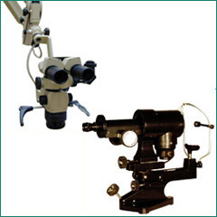
■Manual focusing and
positioning with the help of 320 degree rotatable
counter balanced gas springs loaded arm
■Helps easy variable angle
viewing
■Co axial Illumination done
through fiber optic light guide (cable) from a twin
club, 15V/150W Halogen Lamp light source
■Intensity is continuously
variable from maximum to minimum and vice-versa.
■Provision is available for
photography and CCTV camera (both for monitoring and/or
recording images), assistant's microscope (Ophthalmologic,
ophthalmic surgical microscope,
Operation Microscope,
Ophthalmological
,Ophthalmologyand monocular
observation tube
■Stable floor stand on
five-four castor wheels for easy mobility
■Table mount is also
available instead of floor stand
After much research, we have developed a range of
ophthalmic (ophthalmology) diagnostic equipment. It is specially
designed to meet the varied needs of surgeons across the
world. Our set of diagnostic equipments comprises of the
following:
■Maylite opthalmoscope
■Friston otoscope with 2
aural and 1 nasal specula
■Laryngeal and post-nasal
mirror
■Metal tongue depressor
■Bent arm throat lamp and
spare lamp in a plastic carry case
■Metal handle accommodates
2 C cells |
| Article Source: |
| http://www.indiamart.com/loyalmediopthotech/ophthalmic-instruments.html |
|
Back To Top |
| |
|
Surgical Microscopy and Dentistry |
The concept of
microscopy (Ophthalmologic,
ophthalmic surgical microscope,
Operation Microscope,
Ophthalmological
,Ophthalmology is nearly more than centuries old, but it was
only when advanced technologies were developed that
practicality of the theory had been maximized. No wonder
why the use of surgical operation microscopes (Ophthalmologic,
ophthalmic surgical microscope,
Ophthalmological ,Ophthalmology,Operation
Microscope)in
dentistry is now considered as one of the most effective
and practical technological advancements ever made.
The Value
The importance of high magnification capacity in
dentistry through surgical microscope(Ophthalmologic,
ophthalmic surgical microscope,
Ophthalmological ,Ophthalmology,
Operation Microscope) has been
well known for so many years. But as the years pass by,
the significance of operation microscope(Ophthalmologic,
ophthalmic surgical microscope,
Ophthalmological ,Ophthalmology,Operation
Microscope) in
dentistry has been recognized for more than 20 years in
various fields of dentistry such as restorative
dentistry, periodontics,and endodontics.The very core
of using surgical microscopes (Ophthalmology, Ophthalmological).in dentistry is
its importance as documentation, diagnostic, and patient
education tool.
Reverting back to history, it is a well known fact that surgical microscope (Ophthalmology,
Operation Microscope,Ophthalmologic,
ophthalmic surgical microscope,
Ophthalmological) has already been a
fundamental part of the medical field since its
inception. Surgeries dependent on surgery microscope cannot
proceed because greater magnification of the affected
area can only be seen clearly through a surgical
microscope (Ophthalmology,
Operation Microscope,Ophthalmologic,
ophthalmic surgical microscope,Ophthalmological) .
With this, technologists know that it is important to
magnify certain object especially if it involves
critical observation. And with greater demand for
magnification and illumination, only the surgery microscope is
capable of providing such things. Consequently, experts
in the filed of Dentistry know that with the growing
demand for better quality image, the industry would need
the services of a operating microscope (Ophthalmology,
Operation Microscope,Ophthalmologic,
ophthalmic surgical microscope,
Ophthalmological) to
supply the necessary illumination and magnification.At this point, experts say that even the best vision may
not suffice the required image quality. At some point,
greater magnification and illumination can only be
achieved through operation microscope (Ophthalmology,Ophthalmological).
No wonder why many dentistry schools nowadays are
collaborating with technology to enhance and improve the
growing trend in advanced fields in dentistry. And even
if many people say that the field of dentistry is more
dawdling compared to the other fields in medicine,
still, they are now incorporating such theory into
practical use.Hence, in the field of endodontics, area
of dentistry focusing more on the morphology, diagnosis, and
physiology of periradicular tissues and dental pulp,
surgical microscope (Ophthalmology,
Operation Microscope,Ophthalmologic,
ophthalmic surgical microscope,
Ophthalmological) is commonly used because
experts say that this device improves the visual
perception through greater magnification and effective
illumination.
At some point, people say that using magnification in
the field of dentistry is a sign of weak spot because
magnification was initially associated with aging,
physical abnormality (concerning with sight), concern
for patients who may feel claustrophobic, or simply the
fact that they are now producing great results even
without the use of operating microscopes(Ophthalmology,
Operation Microscope,Ophthalmologic,
ophthalmic surgical microscope,
Ophthalmological).
However, as the years passed by and as technology
continued to propagate the growing world of dentistry,
more and more dentists are incorporating surgical
microscope (Ophthalmology,Ophthalmological) as their primary visual and
documentation tool.
Generally, most dentists employ the services of low
level magnification or loupes. And even if some
restorative dentists may attest that they do not need
high level magnification, still, the fact that they use
magnification for restorative dentistry is a clear proof
that utilization of surgical microscope (Ophthalmology,
Operation Microscope,Ophthalmologic,
ophthalmic surgical microscope,Ophthalmological) cannot
simply be underrated.With this,he utilization and knowledge of operating microscopes
(Ophthalmology, Ophthalmological)as applied in the field
of dentistry is already a requirement for graduation in
most aspiring dentists.
In the field of periodontics, or the study of alveolar
bone, gums, periodontal ligaments, and cementum, or
collectively known as periodontium (supporting structure
of teeth), surgical microscopes (Ophthalmology,
Operation Microscope,Ophthalmologic,
ophthalmic surgical microscope,
Ophthalmological) have taken its
importance by providing support and improvement on the
field of assessment and evaluation. Through this device, periodontics are able to improve their activities with
respect to more delicate handling of tissues, less
patient discomfort, improvement in accuracy, more
predictable functional and esthetic results, and
improved healing times.
Summary
Given all these facts, the primary objective of using operation microscopes (Ophthalmology,
Operation Microscope,Ophthalmologic,
ophthalmic surgical microscope,
Ophthalmological)in the field of dentistry
is to facilitate the procedure using
the device with structures that will not obstruct the
surgeon’s visual capacity while conducting such
operation.
In using surgical microscopes (Operation
Microscope,Ophthalmologic,Ophthalmological,ophthalmic surgical microscope,
Ophthalmology)
in dentistry,
the probabilities of clinical success will be relatively
high. This is because magnification can objectively
improve the visual capacity of the surgeon as well as
refining the images seen and observed. Although some
people may attest that control could be a problem due to
the available angle, it is still important to consider
the different micrology (Ophthalmology,Ophthalmological) techniques that can be
applied so as to simplify the procedure.
Indeed, one cannot simply say that the microscope’s (Ophthalmic
surgical microscope,
Operation Microscope,
Ophthalmologic,
Ophthalmological,Ophthalmology) use in dentistry is just another
development in the making. It is now considered as one
of the most important section in dentistry and is
considered to be very essential in various fields of
dentistry. |
| Article Source: |
| http://www. microscope.com/-microscopy-and-dentistry.html |
|
Back To Top |
| |
|